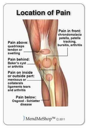
Anatomy of the left knee diagram
Home » Wallpapers » Anatomy of the left knee diagramYour Anatomy of the left knee diagram images are available. Anatomy of the left knee diagram are a topic that is being searched for and liked by netizens now. You can Get the Anatomy of the left knee diagram files here. Download all free photos.
If you’re looking for anatomy of the left knee diagram images information linked to the anatomy of the left knee diagram keyword, you have visit the right site. Our site frequently provides you with hints for seeing the highest quality video and picture content, please kindly search and find more informative video articles and images that match your interests.
Anatomy Of The Left Knee Diagram. It is held in place by a ligament at the bottom and a tendon on top. The pancreas is a glandular organ that produces a number of hormones essential to the body. The part of the knee between the end of the thigh bone femur and the top of the shin bone tibia is called the tibiofemoral joint. The knee joint is made up of two parts.
 Pin Na Doske Chronic Pain From br.pinterest.com
Knee Human Anatomy Function Parts Conditions Treatments This Is Such A Great Diagram Of The Patella My Left Knee Will Knee Joint Picture Image On Medicinenet Com. The anatomy of the knee consists of bones muscles nerves cartilages tendons and ligaments. The kneecap slides along a groove in the femur as the knee bends. The largest joint in the body the knee moves like a hinge allowing you to sit squat walk or jump. The fibula is smaller thinner and laterally positioned compared to the tibia. Ear Anatomy Diagram Labelled Lateral image mediolateral projection of the left knee Latoya Savage Minggu 03 Oktober 2021 In mammals the ear is usually described as having three partsthe outer.
The pancreas is a glandular organ that produces a number of hormones essential to the body.
Those connect to the femur and tibia. The patellofemoral joint is between the end of the thigh bone femur and the kneecap patella. The kneecap slides along a groove in the femur as the knee bends. Therefore it facilitates movement. The MCL is situated within the knee. It is both the longes.
 Source: pinterest.com
The knee is a complex joint that flexes extends and twists slightly from side to side. Twenty years5-9 we have found that its anatomy has only been described qualitatively and there is controversy about descrip-tions of some aspects of its anatomy that have been contra-dictory or incomplete2610-15. Knee Human Anatomy Function Parts Conditions Treatments This Is Such A Great Diagram Of The Patella My Left Knee Will Knee Joint Picture Image On Medicinenet Com. The smaller bone that runs alongside the tibia fibula and the kneecap patella are the other bones that make the knee joint. Anatomy of the Knee.
 Source: pinterest.com
The largest joint in the body the knee moves like a hinge allowing you to sit squat walk or jump. The pelvic bones are smaller and narrower. 11 Diagram Of The Left Knee Anatomy Of The Left Knee Diagram. The medial ligament complex of the knee includes one large ligament and a series of capsular thickenings and tendinous attachments. See knee anatomy stock video clips.
 Source: pinterest.com
Knee Anatomy Diagram. Medial and horizontal guarantee tendons. The knee joint is made up of two parts. Maybe you would like to learn more about one of these. The fibula is smaller thinner and laterally positioned compared to the tibia.
 Source: pinterest.com
Flexion extension medial rotation and lateral rotation and it connects the tibia and the fibula with the thigh bone femur. Labeled Diagram of the Knee Joint. The pelvic bones are smaller and narrower. The major parts of a bird labeled on a real bird photo. Knee joint is one of the most important hinge joints of our body.
 Source: pinterest.com
The pancreas is a glandular organ that produces a number of hormones essential to the body. The medial ligament complex of the knee includes one large ligament and a series of capsular thickenings and tendinous attachments. The kneecap slides along a groove in the femur as the knee bends. Its complexity and its efficiency is the best example of Gods creation. Each of the 6 sections Bones Connective Tissue 1 Connective Tissue 2 Deep Muscles Muscles Skin can be opened up rotated left or right and viewed more closely.
 Source: pinterest.com
Picture of the knee webmd webmd s knee anatomy page provides a detailed image and definition of the knee and its parts including ligaments bones and muscles diagram the left knee anatomy organ diagram the left knee see more about diagram the left knee anatomy of the left knee diagram diagram of back of left knee diagram of left knee. The patellofemoral joint is between the end of the thigh bone femur and the kneecap patella. Its complexity and its efficiency is the best example of Gods creation. Knee Anatomy Diagram. The 3B Scientific Anatomy Video Knee Joint demonstrates the structure of the knee joint.
 Source: pinterest.com
Left Knee Anatomy Diagram. The pelvic bones are smaller and narrower. 11 Diagram Of The Left Knee Anatomy Of The Left Knee Diagram. The knee joins the thigh bone femur to the shin bone. Patella the thick triangular bone that sits over the other bones at the front of the knee or kneecap.
 Source: br.pinterest.com
See knee anatomy stock video clips. Tendons connect the knee bones to. 11 Diagram Of The Left Knee Anatomy Of The Left Knee Diagram. The knee joint is surrounded by synovial fluid which keeps it. The pancreas is a glandular organ that produces a number of hormones essential to the body.
 Source: pinterest.com
Tibia the bone at the front of the lower leg or shin bone. As a result it doesnt play any crucial role in weight bearing. The knee joins the thigh bone femur to the shin bone. Left Knee Anatomy Diagram. The knee consists of three bones.
 Source: pinterest.com
The knee is the largest joint in your body and one of the most easily injuredIt is a pivotal hinge joint in the leg that allows for a variety of movements ie. Labeled structures include the femur patella medial meniscus anterior cruciate ligament or ACL tibia posterior cruciate ligament lateral meniscus and fibula. The knee is a complex joint that flexes extends and twists slightly from side to side. The kneecap slides along a groove in the femur as the knee bends. Our interactive 3D knee diagram is an informative 360 degree rotating model.
 Source: pinterest.com
Twenty years5-9 we have found that its anatomy has only been described qualitatively and there is controversy about descrip-tions of some aspects of its anatomy that have been contra-dictory or incomplete2610-15. Anatomy of the Knee - Medical Illustration Human Anatomy Drawing. The largest joint in the body the knee moves like a hinge allowing you to sit squat walk or jump. The part of the knee between the end of the thigh bone femur and the top of the shin bone tibia is called the tibiofemoral joint. The knee joint is made up of two parts.
 Source: id.pinterest.com
The anatomy of the knee consists of bones muscles nerves cartilages tendons and ligaments. See knee anatomy stock video clips. Patella the thick triangular bone that sits over the other bones at the front of the knee or kneecap. Anatomy of the Knee. The kneecap slides along a groove in the femur as the knee bends.
 Source: pinterest.com
Picture of the knee webmd webmd s knee anatomy page provides a detailed image and definition of the knee and its parts including ligaments bones and muscles diagram the left knee anatomy organ diagram the left knee see more about diagram the left knee anatomy of the left knee diagram diagram of back of left knee diagram of left knee. 37876 knee anatomy stock photos vectors and illustrations are available royalty-free. Twenty years5-9 we have found that its anatomy has only been described qualitatively and there is controversy about descrip-tions of some aspects of its anatomy that have been contra-dictory or incomplete2610-15. The fibula is smaller thinner and laterally positioned compared to the tibia. Maybe you would like to learn more about one of these.
 Source: pinterest.com
The part of the knee between the end of the thigh bone femur and the top of the shin bone tibia is called the tibiofemoral joint. Each of the 6 sections Bones Connective Tissue 1 Connective Tissue 2 Deep Muscles Muscles Skin can be opened up rotated left or right and viewed more closely. The major parts of a bird labeled on a real bird photo. The smaller bone that runs alongside the tibia fibula and the kneecap patella are the other bones that make the knee joint. Patella the thick triangular bone that sits over the other bones at the front of the knee or kneecap.
 Source: pinterest.com
The anatomy of the knee consists of bones muscles nerves cartilages tendons and ligaments. Left Knee Anatomy Diagram. Ear Anatomy Diagram Labelled Lateral image mediolateral projection of the left knee Latoya Savage Minggu 03 Oktober 2021 In mammals the ear is usually described as having three partsthe outer. 37876 knee anatomy stock photos vectors and illustrations are available royalty-free. The medial ligament complex of the knee includes one large ligament and a series of capsular thickenings and tendinous attachments.
 Source: pinterest.com
The kneecap slides along a groove in the femur as the knee bends. Maybe you would like to learn more about one of these. Those connect to the femur and tibia. Patella the thick triangular bone that sits over the other bones at the front of the knee or kneecap. The pancreas is a glandular organ that produces a number of hormones essential to the body.
 Source: pinterest.com
Knee Anatomy Pain Inside. Tendons connect the knee bones to. Left Knee Anatomy Diagram. The pancreas is a glandular organ that produces a number of hormones essential to the body. Its complexity and its efficiency is the best example of Gods creation.
 Source: pinterest.com
37876 knee anatomy stock photos vectors and illustrations are available royalty-free. Patella the thick triangular bone that sits over the other bones at the front of the knee or kneecap. Medial and horizontal guarantee tendons. Ear Anatomy Diagram Labelled Lateral image mediolateral projection of the left knee Latoya Savage Minggu 03 Oktober 2021 In mammals the ear is usually described as having three partsthe outer. The knee is the largest joint in your body and one of the most easily injuredIt is a pivotal hinge joint in the leg that allows for a variety of movements ie.
This site is an open community for users to do sharing their favorite wallpapers on the internet, all images or pictures in this website are for personal wallpaper use only, it is stricly prohibited to use this wallpaper for commercial purposes, if you are the author and find this image is shared without your permission, please kindly raise a DMCA report to Us.
If you find this site value, please support us by sharing this posts to your preference social media accounts like Facebook, Instagram and so on or you can also bookmark this blog page with the title anatomy of the left knee diagram by using Ctrl + D for devices a laptop with a Windows operating system or Command + D for laptops with an Apple operating system. If you use a smartphone, you can also use the drawer menu of the browser you are using. Whether it’s a Windows, Mac, iOS or Android operating system, you will still be able to bookmark this website.