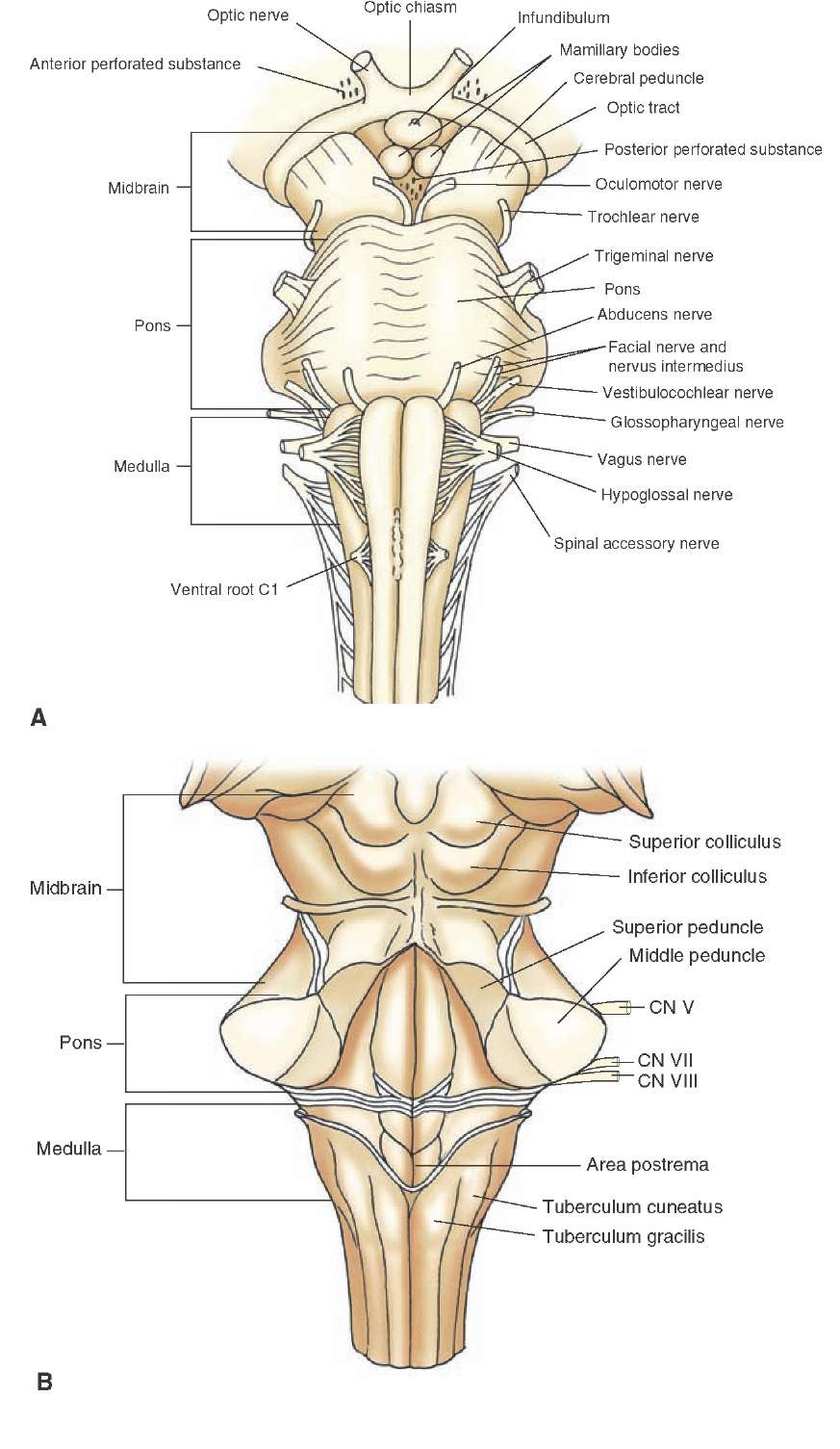
Brainstem diagram
Home » Background » Brainstem diagramYour Brainstem diagram images are available. Brainstem diagram are a topic that is being searched for and liked by netizens today. You can Download the Brainstem diagram files here. Find and Download all free images.
If you’re searching for brainstem diagram pictures information linked to the brainstem diagram keyword, you have come to the right blog. Our site always provides you with suggestions for refferencing the maximum quality video and picture content, please kindly surf and locate more informative video articles and images that match your interests.
Brainstem Diagram. Baseline inhibition inhibition of extemporous movement. This is the largest part of the brain stem. Obviously in individual patients the size of the components of the circle of Willis will. The brainstem is organized.
 Tallo Encefalico Tronco Encefalico Vision Ventral Parte Posterior Del Cerebro Contigua Y Estructural Brain Stem Human Anatomy And Physiology Cranial Nerves From pinterest.com
Many of the most basic survival functions of the brain are controlled by the brainstem. The brainstem is the region of the brain that connects the cerebrum with the spinal cord. Correlation of in vitro mr images with histologic sections 923 mr imaging was performed on three formaldehyde fixed brainstem specimens that were sectioned in the axial plane and myelin stained. Superiorly continuous with forebrain. Axial sections of the brainstem with major. It is the base from which the majority of the cranial nerves arise providing integral sensory information and is critical to our survival in its regulatory role of autonomous bodily functions.
The midbrain or mesencephalon is a very complex structure with a range of different neuron clusters nuclei and colliculi neural pathways and other structures.
If the problem continues send us an email to let us know. The first part of the brainstem we will consider is the midbrain. Midbrain Pons Medulla oblangata. Please try reloading the page. Many of the most basic survival functions of the brain are controlled by the brainstem. The brain stem is located in front of the cerebellum and connects to the spinal cord.
 Source: pinterest.com
Please try reloading the page. The midbrain helps control eye movement and processes visual and. We address two central issues modularity and neural coding by reconstructing and analyzing a wiring diagram from a larval zebrafish brainstem. If the problem continues send us an email to let us know. Posteriorly pons and medulla is seperated by fourth ventricle.
 Source: pinterest.com
Brainstem overview diagram The brainstem begins at the level of the cerebral peduncles anteriorly and the corpora quadrigemina or quadrigeminal plate posteriorly or tectal plate. Through the brainstem terminating in the thalamus. The brainstem is a critical structure of the nervous system functioning as a central point of communication between the central nervous system and the peripheral nervous system. The midbrain or mesencephalon is a very complex structure with a range of different neuron clusters nuclei and colliculi neural pathways and other structures. It continues along a slight posteroinferior course until it ends at the decussation of the pyramids at the level of the foramen magnum of the skull.
 Source: pinterest.com
This is the largest part of the brain stem. SmartDraw includes 1000s of professional healthcare and anatomy chart templates that you can modify and make your own. Embryologically it develops from the mesencephalon and part of the rhombencephalon all of which originate from the neural ectoderm. Create healthcare diagrams like this example called Brainstem in minutes with SmartDraw. These diagrams are based on a collection of resources and are not intended to be used as a definitive reference rather a general guide to the vascular territories.
 Source: pinterest.com
Posteriorly pons and medulla is seperated by fourth ventricle. The brainstem is a critical structure of the nervous system functioning as a central point of communication between the central nervous system and the peripheral nervous system. Brainstem Responsible for automatic survival reflexes Spinal Cord Controls simple reflexes Pathway to neural fibers Medulla Controlsregulates heartbeat and breathing To and from brain Reticular Formation Helps control arousal responds to change in monotony Thalamus Relays sensory information switchboard between sensory neurons and higher. It is intended for the use of medical students working on human anatomy student nurses physiotherapists electro-radiological technicians and residents especially those working in neurology neurosurgery. Brainstem cross-sectional anatomy diagrams Hacking C.
 Source: pinterest.com
It is a connection between the cerebrum the cerebellum and the spinal cord. It acts as a conduit between the forebrain above and the. Brainstem cross-sectional anatomy diagrams. The importance of the brainstem is hard to overstate as many of the key functions of the body are regulated here. This is the largest part of the brain stem.
 Source: br.pinterest.com
Learn vocabulary terms and more with flashcards games and other study tools. It consists of three major parts. Create healthcare diagrams like this example called Brainstem in minutes with SmartDraw. These nuclei each have a specific role. The brainstem is the region of the brain that connects the cerebrum with the spinal cord.
 Source: pinterest.com
Obviously in individual patients the size of the components of the circle of Willis will. Connecting the brain to the spinal cord the brainstem is the most inferior portion of our brain. The brain stem is located in front of the cerebellum and connects to the spinal cord. Its configuration varies at each level but this tract can be seen on all brainstem sections. Brainstem Responsible for automatic survival reflexes Spinal Cord Controls simple reflexes Pathway to neural fibers Medulla Controlsregulates heartbeat and breathing To and from brain Reticular Formation Helps control arousal responds to change in monotony Thalamus Relays sensory information switchboard between sensory neurons and higher.
 Source: pinterest.com
We address two central issues modularity and neural coding by reconstructing and analyzing a wiring diagram from a larval zebrafish brainstem. The brainstem a posterior structure of the brain comprises the medulla oblongata the pons and the midbrain. It acts as a conduit between the forebrain above and the. Obviously in individual patients the size of the components of the circle of Willis will. Hypertonic-hypokinetic and hypotonic-hyperkinetic syndromes.
 Source: pinterest.com
Correlation of in vitro mr images with histologic sections 923 mr imaging was performed on three formaldehyde fixed brainstem specimens that were sectioned in the axial plane and myelin stained. The importance of the brainstem is hard to overstate as many of the key functions of the body are regulated here. Connecting the brain to the spinal cord the brainstem is the most inferior portion of our brain. Midbrain pons and medulla connected to cerebellum by superiormiddle and inferior cerebellar peduncle resp. These diagrams are based on a collection of resources and are not intended to be used as a definitive reference rather a general guide to the vascular territories.
 Source: pinterest.com
Its configuration varies at each level but this tract can be seen on all brainstem sections. It is intended for the use of medical students working on human anatomy student nurses physiotherapists electro-radiological technicians and residents especially those working in neurology neurosurgery. We identified a recurrently connected center within the 3000-node graph and applied graph clustering algorithms to divide the center into two modules with stronger connectivity within than between modules. Its configuration varies at each level but this tract can be seen on all brainstem sections. It acts as a conduit between the forebrain above and the.
 Source: pinterest.com
The first part of the brainstem we will consider is the midbrain. Nigrostriatal and thalamostriatal pathways. This human anatomy module is about the cranial nerves. We identified a recurrently connected center within the 3000-node graph and applied graph clustering algorithms to divide the center into two modules with stronger connectivity within than between modules. Baseline inhibition inhibition of extemporous movement.
 Source: pinterest.com
The midbrain helps control eye movement and processes visual and. The cochlear nucleus the superior olivary complex the lateral lemniscus and the. The brainstem is a critical structure of the nervous system functioning as a central point of communication between the central nervous system and the peripheral nervous system. This human anatomy module is about the cranial nerves. Its configuration varies at each level but this tract can be seen on all brainstem sections.
 Source: pinterest.com
Anatomy and physiology of brain stem. And the rest contain both afferent and efferent fibers the trigeminal nerve facial nerve glossopharyngeal nerve and. The brainstem is a critical structure of the nervous system functioning as a central point of communication between the central nervous system and the peripheral nervous system. Most cranial nerves are found in the brainstem. The brainstem a posterior structure of the brain comprises the medulla oblongata the pons and the midbrain.
 Source: pinterest.com
Through the brainstem terminating in the thalamus. The brainstem is a critical structure of the nervous system functioning as a central point of communication between the central nervous system and the peripheral nervous system. The brainstem is composed of the midbrain the pons and the medulla oblongata situated in the posterior part of the brain. Hypertonic-hypokinetic and hypotonic-hyperkinetic syndromes. Anatomy and physiology of brain stem.
 Source: pinterest.com
The brainstem is composed of the midbrain the pons and the medulla oblongata situated in the posterior part of the brain. Superiorly continuous with forebrain. This is the largest part of the brain stem. The brainstem is made of three. Brainstem cross-sectional anatomy diagrams Hacking C.
 Source: pinterest.com
The first part of the brainstem we will consider is the midbrain. Most cranial nerves are found in the brainstem. Brainstem overview diagram The brainstem begins at the level of the cerebral peduncles anteriorly and the corpora quadrigemina or quadrigeminal plate posteriorly or tectal plate. SmartDraw includes 1000s of professional healthcare and anatomy chart templates that you can modify and make your own. It consists of three major parts.
 Source: pinterest.com
The first part of the brainstem we will consider is the midbrain. It continues along a slight posteroinferior course until it ends at the decussation of the pyramids at the level of the foramen magnum of the skull. The auditory pathway like that of other senses except for smell and sight passes through a number of relay stations within the brainstem. The brain stem is located in front of the cerebellum and connects to the spinal cord. Brainstem Responsible for automatic survival reflexes Spinal Cord Controls simple reflexes Pathway to neural fibers Medulla Controlsregulates heartbeat and breathing To and from brain Reticular Formation Helps control arousal responds to change in monotony Thalamus Relays sensory information switchboard between sensory neurons and higher.
 Source: pinterest.com
Through the brainstem terminating in the thalamus. The brainstem a posterior structure of the brain comprises the medulla oblongata the pons and the midbrain. Learn vocabulary terms and more with flashcards games and other study tools. Brainstem overview diagram The brainstem begins at the level of the cerebral peduncles anteriorly and the corpora quadrigemina or quadrigeminal plate posteriorly or tectal plate. It is a connection between the cerebrum the cerebellum and the spinal cord.
This site is an open community for users to share their favorite wallpapers on the internet, all images or pictures in this website are for personal wallpaper use only, it is stricly prohibited to use this wallpaper for commercial purposes, if you are the author and find this image is shared without your permission, please kindly raise a DMCA report to Us.
If you find this site serviceableness, please support us by sharing this posts to your own social media accounts like Facebook, Instagram and so on or you can also bookmark this blog page with the title brainstem diagram by using Ctrl + D for devices a laptop with a Windows operating system or Command + D for laptops with an Apple operating system. If you use a smartphone, you can also use the drawer menu of the browser you are using. Whether it’s a Windows, Mac, iOS or Android operating system, you will still be able to bookmark this website.