Chambers of heart diagram
Home » Wallpapers » Chambers of heart diagramYour Chambers of heart diagram images are ready in this website. Chambers of heart diagram are a topic that is being searched for and liked by netizens now. You can Find and Download the Chambers of heart diagram files here. Download all royalty-free photos.
If you’re searching for chambers of heart diagram images information connected with to the chambers of heart diagram topic, you have visit the ideal blog. Our website always provides you with hints for seeing the maximum quality video and image content, please kindly surf and locate more informative video articles and graphics that match your interests.
Chambers Of Heart Diagram. The heart is divided into two chambers left and right the right atrium and ventricle lie on the right side and the left atrium and ventricle on the left side. This covering is like a membrane which holds all the parts of the heart. The heart has four chambers. Function and anatomy of the heart made easy using labeled diagrams of cardiac.
 How To Draw Internal Structure Of Human Heart Easy Version Simple Heart Diagram Human Heart Diagram Heart Diagram From pinterest.com
The heart has four chambers two relatively small upper chambers called atria and two larger lower chambers called ventricles. Synchronization of the Two Chamber. The bottom line represents the first and second heart sounds. This covering is like a membrane which holds all the parts of the heart. The boxes are numbered to correlate with the labeled chambers on the cartoon diagram. The right side of the heart has less myocardium in its walls than the left side because the left side has to pump blood through the entire body while the right side only has to pump to the lungs.
Among which the right atrium and ventricle make up the right portion of the heart and the left atrium and ventricle make up the left portion of the heart.
The right atrium left atrium right ventricle and left ventricle. Innermost layer of hearts tissue lines chambers. Exterior of the Human Heart. Red arrows oxygenated blood. The wall of the heart has three different layers such as the Myocardium the Epicardium and the Endocardium. The heart is made up of two chambers.
 Source: id.pinterest.com
13 Heart Chambers And Valves Diagram. Diagram of a heart detailing all major sections. The upper two chambers of the heart are called auricles. 13 Heart Chambers And Valves Diagram. The right ventricle receives blood from the right atrium and pumps it to the lungs.
 Source: nl.pinterest.com
Stud the diagram carefully and answer the questions that follow. The right ventricle receives blood from the right atrium and pumps it to the lungs. The right and left side or chambers of the heart work in tandem with each other. The bottom line represents the first and second heart sounds. Well-Labelled Diagram of Heart.
 Source: pinterest.com
The heart valves which keep blood flowing in the right direction are gates at the chamber openings for the tricuspid and mitral valves and. Synchronization of the Two Chamber. Like the brain the heart continues working tirelessly without stopping for a second and it makes the blood flow through all the blood vessels in your body in one minute. The outer layer of the heart wall is the epicardium the middle layer is the myocardium and the inner layer is the endocardium. This covering is like a membrane which holds all the parts of the heart.
 Source: pinterest.com
Among which the right atrium and ventricle make up the right portion of the heart and the left atrium and ventricle make up the left portion of the heart. It consist of four chambers four valves arteries named as coronary arteries and the conduction system. Separation of right and left side of heart allows efficient supply. Heres more about these three layers. The right atrium left atrium right ventricle and left ventricle.
 Source: pinterest.com
The internal cavity of the heart is divided into four chambers. Well-Labelled Diagram of Heart. The middle layer of the heart wall is called myocardium. Two atria right and left and two ventricles right and left. All these chambers are separated by a layer of tissues known as septum.
 Source: pinterest.com
The boxes are numbered to correlate with the labeled chambers on the cartoon diagram. Right atrium and ventricle of the heart labeled. The heart is divided into two chambers left and right the right atrium and ventricle lie on the right side and the left atrium and ventricle on the left side. Chambers of the Heart. The two atria are thin-walled chambers that receive blood from the veins.
 Source: za.pinterest.com
The left ventricle has about three time the mucsle than the right ventricle. Outermost layer of the heart it is a double-walled sac that encloses the heart. Well-Labelled Diagram of Heart. The ventricles are the chambers that pump blood and atrium are the chambers that receive the blood. The heart valves which keep blood flowing in the right direction are gates at the chamber openings for the tricuspid and mitral valves and.
 Source: pinterest.com
Separation of right and left side of heart allows efficient supply. The image above shows the four chambers of the heart along with major blood vessels and valves. Diagram the anatomical structure of the heart. Synchronization of the Two Chamber. Well-Labelled Diagram of Heart.
 Source: pinterest.com
Red arrows oxygenated blood. Separation of right and left side of heart allows efficient supply. The heart wall is made up of three layers. Blood drains into the atria from the pulmonary and systemic circulatory systems. A summary the heart is located in the thoracic cavity the heart has.
 Source: in.pinterest.com
Now that we have a good understanding of the blood flow through the heart using the cartoon diagrams we can apply it to a more realistic image of the heart. Blue arrows deoxygenated blood. The following diagram shows blood flow through the heart. A powerful natural pump in your body the heart uses its chambers to manage the flow of blood in two distinct circuits ie. Easy steps to draw human heart class 10 ncert write down each step with hand drawn labelled diagram.
 Source: pinterest.com
The heart is made up of two chambers. The wall of the heart has three different layers such as the Myocardium the Epicardium and the Endocardium. The hearts four chambers are. The heart valves which keep blood flowing in the right direction are gates at the chamber openings for the tricuspid and mitral valves and. Diagram the anatomical structure of the heart.
 Source: pinterest.com
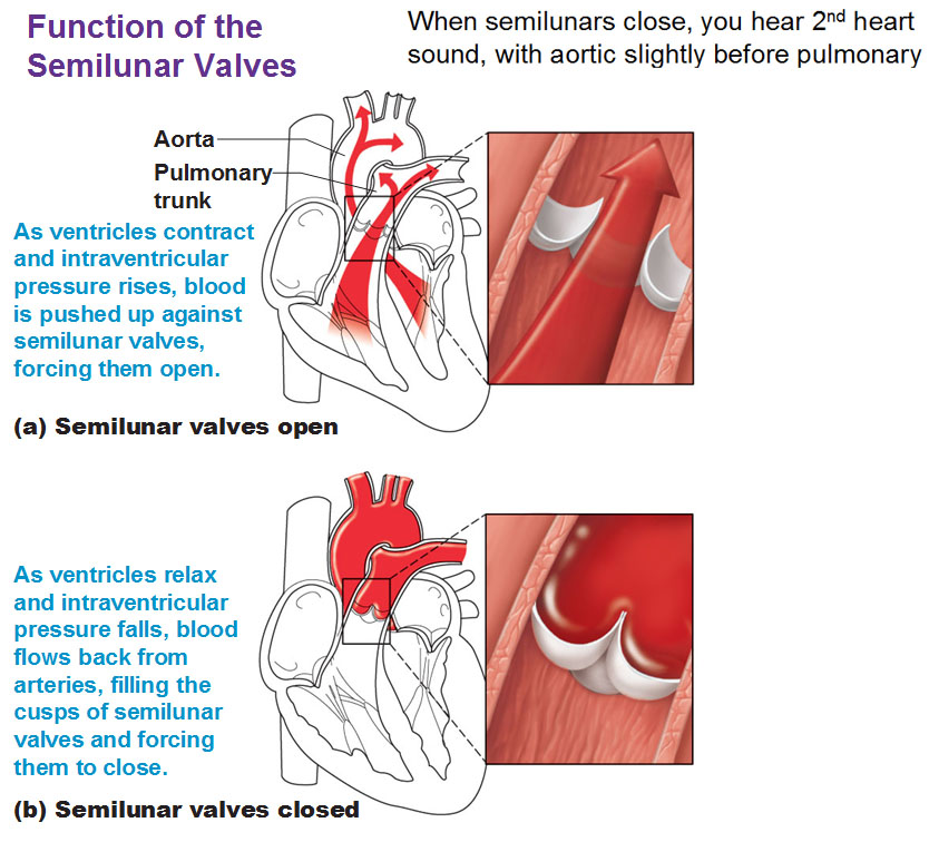
13 Heart Chambers And Valves Diagram. These pressure changes result in the movement of blood through different chambers of the heart and the body as a whole. The heart consist of 4 chambers-2 atria and 2 ventricle. The right ventricle receives blood from the right atrium and pumps it to the lungs. The right side of the heart has less myocardium in its walls than the left side because the left side has to pump blood through the entire body while the right side only has to pump to the lungs.
 Source: pinterest.com
The upper two chambers of the heart are called auricles. These pressure changes result in the movement of blood through different chambers of the heart and the body as a whole. The atria is about 13 the size of the ventricle. 13 Heart Chambers And Valves Diagram. The ventricles are the chambers that pump blood and atrium are the chambers that receive the blood.
 Source: pinterest.com
These pressure changes result in the movement of blood through different chambers of the heart and the body as a whole. It consist of four chambers four valves arteries named as coronary arteries and the conduction system. The lower two chambers of the heart are called ventricles. These pressure changes result in the movement of blood through different chambers of the heart and the body as a whole. The ventricles are the chambers that pump blood and atrium are the chambers that receive the blood.
 Source: pinterest.com
Inside the heart is divided into four heart chambers. Diagram of a heart detailing all major sections. The atria are smaller than the ventricles and have thinner less muscular walls than the. The upper chambers the right and left atria receive incoming blood. Separation of right and left side of heart allows efficient supply.
 Source: pinterest.com
The atria is about 13 the size of the ventricle. A powerful natural pump in your body the heart uses its chambers to manage the flow of blood in two distinct circuits ie. Heart functionally can be separated in left and right side. The upper chambers the right and left atria receive incoming blood. Exterior of the Human Heart.
This site is an open community for users to do sharing their favorite wallpapers on the internet, all images or pictures in this website are for personal wallpaper use only, it is stricly prohibited to use this wallpaper for commercial purposes, if you are the author and find this image is shared without your permission, please kindly raise a DMCA report to Us.
If you find this site convienient, please support us by sharing this posts to your own social media accounts like Facebook, Instagram and so on or you can also bookmark this blog page with the title chambers of heart diagram by using Ctrl + D for devices a laptop with a Windows operating system or Command + D for laptops with an Apple operating system. If you use a smartphone, you can also use the drawer menu of the browser you are using. Whether it’s a Windows, Mac, iOS or Android operating system, you will still be able to bookmark this website.