Dermis diagram
Home » Wallpapers » Dermis diagramYour Dermis diagram images are available. Dermis diagram are a topic that is being searched for and liked by netizens now. You can Get the Dermis diagram files here. Get all royalty-free vectors.
If you’re looking for dermis diagram pictures information related to the dermis diagram interest, you have visit the ideal site. Our website frequently provides you with hints for seeking the highest quality video and picture content, please kindly hunt and locate more informative video articles and images that fit your interests.
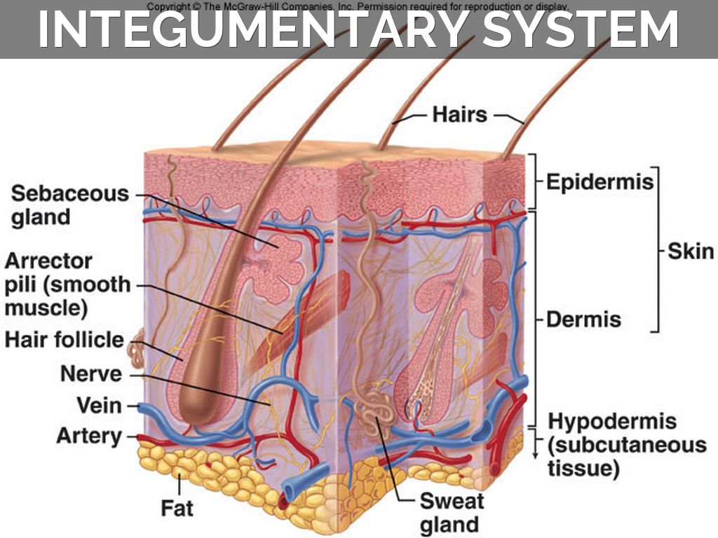
Dermis Diagram. The central pale type A nests are contrasted to the neighboring type B nests. This diagram shows the layers found in skin. INTEGUMENTARY SYSTEM PART III. There are three main layers.
 Labeled Skin Diagrams Health Pictures Skin Structure Skin Anatomy Integumentary System From pinterest.com
It is made up of three layers the epidermis dermis. An example is the skin on the. Download scientific diagram the skin model used for modeling contains seven layers. Skin is the largest organ in the body and covers the bodys entire external surface. There are 3 layers of the skin the epidermis dermis and subcutaneous tissue. Which is the thickest layer.
ACCESSORY STRUCTURES Integumentary Accessory Structures Hair hair follicles sebaceous glands sweat glands and nails.
The junction between the epidermis and dermis of skin histology is known as the dermal ridge or papillae. Is the innermost layer of the skin and is mainly composed of fat and connective tissue. Which is the thickest layer. This diagram shows the layers found in skin. It is made up of three layers the epidermis dermis. Another example biopsy of eyelid skin shows type A and type B nests.
 Source: pinterest.com
As you can see in the skin diagram many structures are embedded in the dermis. Add the following labels to the diagram of the skin shown below. The dermis is a connective tissue layer sandwiched between the epidermis and subcutaneous tissue. Try these curated collections. They are also involved in regulating body temperature.
 Source: pinterest.com
The dermis is composed of two layers. Dermis or corium layer The dermis is a tough and elastic layer containing white fibrous tissue interlaced with yellow elastic fibers. The dermis is the layer of the skin present beneath the epidermis of the skin. 18507 dermis stock photos vectors and illustrations are available royalty-free. An example is the skin on the.
 Source: pinterest.com
While the epidermis covers your body. Epidermis dermis fat cells hair shaft hair follicle hair erector muscle sweat gland pore of sweat gland sebaceous gland blood. While the epidermis covers your body. Which is the thickest layer. The melanophages usually accompany the type A nests.
 Source: pinterest.com
The dermis is a connective tissue layer sandwiched between the epidermis and subcutaneous tissue. Add the following labels to the diagram of the skin shown below. The central pale type A nests are contrasted to the neighboring type B nests. It is the layer of skin you touch when buying any leather goods. Try these curated collections.
 Source: pinterest.com
Download scientific diagram the skin model used for modeling contains seven layers. Download scientific diagram the skin model used for modeling contains seven layers. The epidermis made of closely packed epithelial cells and the dermis made of dense. Hair follicle microscope layers of dermis skin istology structure of dermis structure skin the skin anatomy the structure of the skin subcutaneous layer the skin an layersskin diagrams. The dermis is also involved in the synthesis of Vitamin D on exposure to sunlight.
 Source: id.pinterest.com
Type A nests have gray cytoplasm with hyperchromatic smudged nuclei. Skin is the largest organ in the body and covers the bodys entire external surface. The dermis is a tough layer of skin. The dermis the epidermis fat layer 2. It is made up of three layers the epidermis dermis.
 Source: pinterest.com
The dermis is the layer of the skin present beneath the epidermis of the skin. The epidermis made of closely packed epithelial cells and the dermis made of dense. The deep papillary dermis has a smooth round outline with proper maturation even at this power. 18507 dermis stock photos vectors and illustrations are available royalty-free. There are 3 layers of the skin the epidermis dermis and subcutaneous tissue.
 Source: pinterest.com
Try these curated collections. There are 3 layers of the skin the epidermis dermis and subcutaneous tissue. The outermost layer of the skin is. Full color diagram of skin anatomy. Try these curated collections.
 Source: pinterest.com
Lying underneath the epidermisthe most superficial layer of our skinis the dermis sometimes called the corium. Humans shed around 500 million skin cells each day. We are pleased to provide you with the picture named Epidermis Dermis Anatomical Location Diagram. While the epidermis covers your body. The dermis the epidermis.
 Source: pinterest.com
Type A nests have gray cytoplasm with hyperchromatic smudged nuclei. The outermost layer of the skin is. The melanophages usually accompany the type A nests. This diagram shows the layers found in skin. Try these curated collections.
 Source: pinterest.com
Start studying Epidermis Dermis Label Quiz. The dermis is split into two parts. The skins structure is made up of an intricate network which serves as the bodys initial barrier against pathogens UV light and chemicals and mechanical injury. The dermis is a tough layer of skin. Tips and advice on skin.
 Source: pinterest.com
The skins structure is made up of an intricate network which serves as the bodys initial barrier against pathogens UV light and chemicals and mechanical injury. Hair follicle microscope layers of dermis skin istology structure of dermis structure skin the skin anatomy the structure of the skin subcutaneous layer the skin an layersskin diagrams. Lying underneath the epidermisthe most superficial layer of our skinis the dermis sometimes called the corium. The subcutaneous layer under the dermis is made up of connective tissue and fat a good insulator. Add the following labels to the diagram of the skin shown below.
 Source: pinterest.com
The melanophages usually accompany the type A nests. Try these curated collections. There are three main layers. Now lets known about the histology of the dermis of animals. The subcutaneous layer under the dermis is made up of connective tissue and fat a good insulator.
 Source: pinterest.com
It is made up of three layers the epidermis dermis. Epidermis dermis fat cells hair shaft hair follicle hair erector muscle sweat gland pore of sweat gland sebaceous gland blood. Which is the thickest layer. Find normal skin anatomy stock images in hd and millions of other. The dermis is also involved in the synthesis of Vitamin D on exposure to sunlight.
 Source: id.pinterest.com
The dermis is a tough layer of skin. Download scientific diagram the skin model used for modeling contains seven layers. 18507 dermis stock photos vectors and illustrations are available royalty-free. An example is the skin on the. The dermis is split into two parts.
 Source: pinterest.com
This layer constitutes of fat fibres collagen and blood vessels which make the skin flexible and strong. Tips and advice on skin. The melanophages usually accompany the type A nests. Epidermis dermis fat cells hair shaft hair follicle hair erector muscle sweat gland pore of sweat gland sebaceous gland blood. Learn vocabulary terms and more with flashcards games and other study tools.
 Source: pinterest.com
It is made up of three layers the epidermis dermis and the hypodermis all three of which vary significantly in their anatomy and function. The dermis or corium is a layer of skin between the epidermis and subcutaneous tissues that primarily consists of dense irregular connective tissue and cushions the body from stress and strain. Now lets known about the histology of the dermis of animals. The dermis is composed of two layers. The dermis is split into two parts.
 Source: pinterest.com
See dermis stock video clips. The dermis is also involved in the synthesis of Vitamin D on exposure to sunlight. Now lets known about the histology of the dermis of animals. INTEGUMENTARY SYSTEM PART III. Male Digestive System Diagram 2021 Male and Female Digestive system anatomy and physiology.
This site is an open community for users to do sharing their favorite wallpapers on the internet, all images or pictures in this website are for personal wallpaper use only, it is stricly prohibited to use this wallpaper for commercial purposes, if you are the author and find this image is shared without your permission, please kindly raise a DMCA report to Us.
If you find this site helpful, please support us by sharing this posts to your favorite social media accounts like Facebook, Instagram and so on or you can also bookmark this blog page with the title dermis diagram by using Ctrl + D for devices a laptop with a Windows operating system or Command + D for laptops with an Apple operating system. If you use a smartphone, you can also use the drawer menu of the browser you are using. Whether it’s a Windows, Mac, iOS or Android operating system, you will still be able to bookmark this website.