Diagram of cranium
Home » Background » Diagram of craniumYour Diagram of cranium images are ready. Diagram of cranium are a topic that is being searched for and liked by netizens now. You can Find and Download the Diagram of cranium files here. Get all free photos and vectors.
If you’re looking for diagram of cranium pictures information connected with to the diagram of cranium interest, you have visit the right site. Our site frequently gives you suggestions for seeing the maximum quality video and picture content, please kindly search and find more informative video articles and graphics that match your interests.
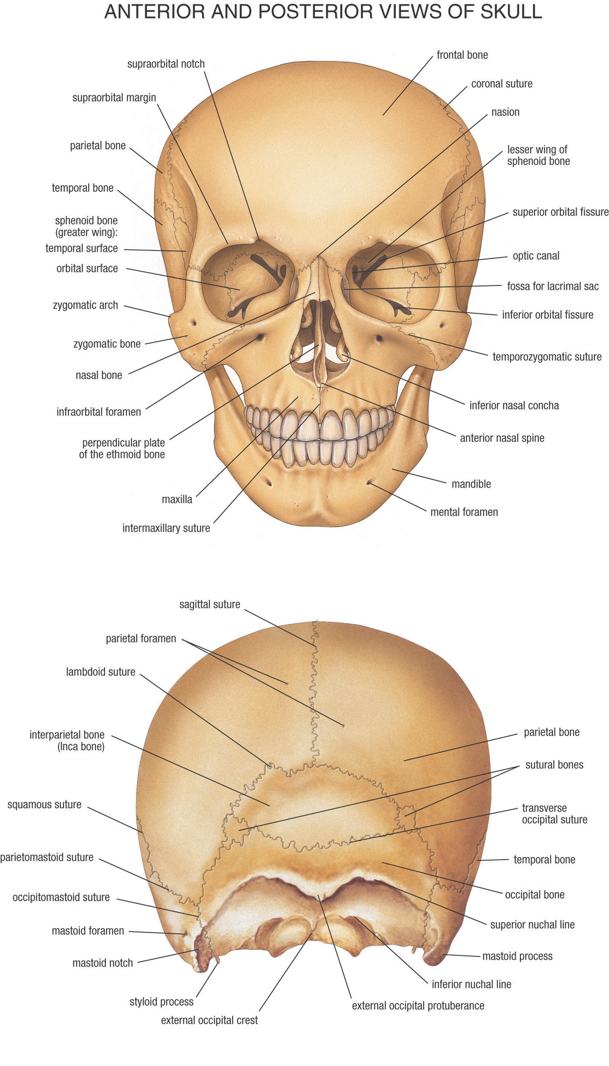
Diagram Of Cranium. The cranial bones compose the top and back of the skull and enclose the brain. The cranial nerves are named after the body parts that they serve and are also assigned Roman numerals based off their location from front to back. The skull consists of the cranial bones and the facial skeleton. Explore the interactive 3-D diagram below to learn more about the cranial bones.
 Parts Of The Human Skull Biology101 Study Guide Anatomy And Physiology Human Anatomy And Physiology Physiology From pinterest.com
It controls the eyes lateral rectus muscle which moves the eye sideways away from the nose. Oh Oh Oh To Touch And Feel Very Good Velvet such-A Heaven. Like spinal nerves cranial nerves are bundles of sensory and motor neurons that conduct. The cranium skull is the skeletal structure of the head that supports the face and protects the brain. And the rest contain both afferent and efferent fibers the trigeminal nerve facial. The 14 bones of the facial skeleton form the entrances to the respiratory and digestive tracts.
The cranial floor consists of the sphenoid and ethmoid bones.
Mnemonic for Function of Cranial Nerves. It is subdivided into the facial bones and the brain caseSkull anatomy unlabeled In this image you will find Skull anatomy in it. See if you can identify the different cranial bones that are labeled in the cranium bone quiz below. The cranium is made up of 8 bones. Anatomically the cranium can be subdivided into a roof and a base. Facial Aspect of Cranium Features of the anterior or facial frontal aspect of the cranium are.
 Source: pinterest.com
The frontal bone specifically its squamous flat part forms the skeleton of the forehead articulating inferiorly with the nasal and zygomatic bones. Like spinal nerves cranial nerves are bundles of sensory and motor neurons that conduct. Diagrams of cranial nerves. The eight major bones of the cranium are connected by cranial sutures which are fibrous bands of tissue that resemble seams. Cranial bone conditions Several injuries and health conditions can impact your cranial bones including fractures.
 Source: pinterest.com
Often the ossicles of the ear and the hyoid bone are counted as part of the skull. This is a diagram of a skull that displays the components of neurocranium. Both the liver and the stomach are located in the lower chest region under the thoracic diaphragm a sheet of muscle at the bottom of the rib cage that separates the chest cavity from the abdominal cavity. Mnemonic for Function of Cranial Nerves. The Mandible is the only bone in the skull that can move.
 Source: pinterest.com
Oh Oh Oh To Touch And Feel Very Good Velvet such-A Heaven. Facial Aspect of Cranium Features of the anterior or facial frontal aspect of the cranium are. The eight major bones of the cranium are connected by cranial sutures which are fibrous bands of tissue that resemble seams. This is a diagram of a skull that displays the components of neurocranium. Learn the major cranial bone names and anatomy of the skull using this mnemonic and labeled diagram.
 Source: pinterest.com
These original anatomical drawings were produced digitally working from medical imaging sources and 3D reconstructions using Adobe Illustrator. Sutures connect cranial bones and facial bones of the skull. Like spinal nerves cranial nerves are bundles of sensory and motor neurons that conduct. The remainder of the bones in the skull are the facial bones. Five are efferent the oculomotor nerve trochlear nerve abducent nerve accessory nerve and hypoglossal nerve.
 Source: pinterest.com
Download scientific diagram diagram of the 12 cranial nerves and their functional connections. Sutures connect cranial bones and facial bones of the skull. It encloses and protects the brain meninges and cerebral vasculature. Cranial nerves are either efferent afferent or both. Bones of the Cranium Diagram Quizlet.
 Source: pinterest.com
There are 12 pairs of cranial nerves attached to the brain. The skull consists of the cranial bones and the facial skeleton. The principal bones that form the cranium are the occipital bone behind and below the parietal bone and temporal bone on each side the sphenoid bone. Facial Aspect of Cranium Features of the anterior or facial frontal aspect of the cranium are. Explore the interactive 3-D diagram below to learn more about the cranial bones.
 Source: pinterest.com
The eight major bones of the cranium are connected by cranial sutures which are fibrous bands of tissue that resemble seams. Anatomically the cranium can be subdivided into a roof and a base. The skull consists of the cranial bones and the facial skeleton. The cranium is the part of the skull that protects the brain and it is made up of eight bones and it also supports the structure of the face. It makes up the roof of the eye orbits.
 Source: pinterest.com
There are eight major bones and eight auxiliary bones of the cranium. A diagram of the human skeleton showing bone and cartilage. The cranial floor consists of the sphenoid and ethmoid bones. Develop a good way to remember the cranial bone markings types definition and names including the frontal bone occipital bone parietal bone temporal bone sphenoid bone and ethmoid bone using this. It is also known as the calvarium.
 Source: pinterest.com
Download scientific diagram diagram of the 12 cranial nerves and their functional connections. Both the liver and the stomach are located in the lower chest region under the thoracic diaphragm a sheet of muscle at the bottom of the rib cage that separates the chest cavity from the abdominal cavity. Diagrams of cranial nerves. The facial skeleton is formed by the mandible maxillae. Explore the interactive 3-D diagram below to learn more about the cranial bones.
 Source: pinterest.com
Learn here the cavities of the human body. Five are efferent the oculomotor nerve trochlear nerve abducent nerve accessory nerve and hypoglossal nerve. The facial skeleton is referred to as. Often the ossicles of the ear and the hyoid bone are counted as part of the skull. See if you can identify the different cranial bones that are labeled in the cranium bone quiz below.
 Source: pinterest.com
The central nervous system lies largely within the axial skeleton the brain being well protected by the cranium and the spinal cord by the vertebral column by means of the bony neural arches the arches of bone that encircle the spinal cord and the intervening ligaments. The Mandible is the only bone in the skull that can move. Figure 67 and Figure 68 show all the bones of the skull as they appear from the outside. The facial area includes the zygomatic or malar. The cranial bones compose the top and back of the skull and enclose the brain.
 Source: pinterest.com
They protect and surround the brain. Download scientific diagram schematic drawing showing connections between cranial nerves vii ix and x and some of their branches. In humans the base of the cranium is the occipital bone which has a central opening foramen magnum to admit the spinal cordThe parietal and temporal bones form the sides and uppermost portion of the dome of the cranium and the frontal bone forms the forehead. Figure 67 and Figure 68 show all the bones of the skull as they appear from the outside. There are eight major bones and eight auxiliary bones of the cranium.
 Source: pinterest.com
Learn here the cavities of the human body. The skull base comprises parts of the frontal ethmoid sphenoid occipital and temporal bones. The following anatomy diagrams of the skull contain the parts of bones and lateral view of the skull. Like spinal nerves cranial nerves are bundles of sensory and motor neurons that conduct. The cranial bones compose the top and back of the skull and enclose the brain.
 Source: pinterest.com
The following anatomy diagrams of the skull contain the parts of bones and lateral view of the skull. The facial skeleton as its name suggests makes up the face of the skull. Our cranial bones do not fuse but remain distinct and separate throughout our lives. Like spinal nerves cranial nerves are bundles of sensory and motor neurons that conduct. Looking at it from the inside it can be subdivided into the anterior middle and posterior cranial fossae.
 Source: pinterest.com
These original anatomical drawings were produced digitally working from medical imaging sources and 3D reconstructions using Adobe Illustrator. The cranial nerves are named after the body parts that they serve and are also assigned Roman numerals based off their location from front to back. The following anatomy diagrams of the skull contain the parts of bones and lateral view of the skull. The cervical nerves consist of eight paired nerves. The occipital bone forms the base of the skull at the rear of the cranium.
 Source: pinterest.com
The occipital is cupped like a saucer in. The facial area includes the zygomatic or malar. The cranium skull is the skeletal structure of the head that supports the face and protects the brain. There are eight major bones and eight auxiliary bones of the cranium. Explore the interactive 3-D diagram below to learn more about the cranial bones.
 Source: pinterest.com
It makes up the roof of the eye orbits. In most people the cranium is made up of 8 bones and the face is made up of 14. The following anatomy diagrams of the skull contain the parts of bones and lateral view of the skull. It is also known as the calvarium. The cranial nerves are a set of twelve nerves that originate in the brain.
 Source: pinterest.com
The cranium skull is the skeletal structure of the head that supports the face and protects the brain. This diagram depicts human body map of organs with parts and labels. Five are efferent the oculomotor nerve trochlear nerve abducent nerve accessory nerve and hypoglossal nerve. Only one of those that stem from the brainstem is afferent the vestibulocochlear nerve. The cervical nerves consist of eight paired nerves.
This site is an open community for users to share their favorite wallpapers on the internet, all images or pictures in this website are for personal wallpaper use only, it is stricly prohibited to use this wallpaper for commercial purposes, if you are the author and find this image is shared without your permission, please kindly raise a DMCA report to Us.
If you find this site value, please support us by sharing this posts to your preference social media accounts like Facebook, Instagram and so on or you can also bookmark this blog page with the title diagram of cranium by using Ctrl + D for devices a laptop with a Windows operating system or Command + D for laptops with an Apple operating system. If you use a smartphone, you can also use the drawer menu of the browser you are using. Whether it’s a Windows, Mac, iOS or Android operating system, you will still be able to bookmark this website.