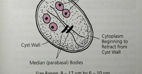
Diagram of entamoeba histolytica
Home » Wallpapers » Diagram of entamoeba histolyticaYour Diagram of entamoeba histolytica images are available. Diagram of entamoeba histolytica are a topic that is being searched for and liked by netizens now. You can Download the Diagram of entamoeba histolytica files here. Download all royalty-free images.
If you’re looking for diagram of entamoeba histolytica images information related to the diagram of entamoeba histolytica topic, you have come to the ideal blog. Our site always provides you with suggestions for seeing the highest quality video and picture content, please kindly search and find more enlightening video content and images that match your interests.
Diagram Of Entamoeba Histolytica. Iv In the nucleus a peripheral ring of granule. It may occur in the liver and lungs. Entamoeba histolytica is a parasite and lives in the mucous and sub-mucous layers of the large intestine of man. Entamoeba Histolytica is an infectious parasite found in the human intestine and several other primates.
 Intestinal Amebae Subphylum Sarcodina Phylum Sarcomastigophora Iodamoeba Butschii Cyst Ch Medical Laboratory Science Med School Motivation School Stickers From pinterest.com
Entamoeba histolytica is a common protozoan parasite found in the large intestine of human. Entamoeba histolytica is a parasite and lives in the mucous and sub-mucous layers of the large intestine of man. Details and draw labelled diagrams of the pathogens. Exercise 14 Observation A. It was discovered by a Russian zoologist Friedrick Losch in 1875. The genus Entamoeba was established by Leidy in 1879.
Histolytica is a microscopic endoparasite and is the causative agent of amoebic dysentery in man.
The disease caused by the parasite is known as Amoebic dysentery. Rarely it invades brain spleen etc. Entos - within. Entamoeba Observe the following features of the parasite in the slide or photograph. Exercise 14 Observation A. Entamoeba histolytica is one of a number of species of small amoebae which live in the alimentary canal of humans.
 Source: pinterest.com
Entos - within. Entamoeba histolytica are pathogenic amoeba that are widely known for causing intestinal and extraintestinal infections in human beings. A constriction appears in the middle which grows deeper and the nucleus is divided into two by a modified type of mitosis. Entos - within. Eh is an anaerobic amoebozoan an extracellular enteric parasite and amongst the most pathogenic protozoans.
 Source: pinterest.com
Histolytica is estimated to infect about 35-50 million people worldwide. It is cosmopolitan in distribution but more common in the tropical and subtropical regions of the world. Entamoeba histolytica is an anaerobic parasitic amoebozoan part of the genus Entamoeba. The parasite is responsible for amoebiasis and liver absceses. As a result it has the capability of causing critical diseases like amoebiasis or amoebic dysentery.
 Source: pinterest.com
Histolytica is estimated to infect about 35-50 million people worldwide. In most infected individuals the trophozoites exist as commensals. 185 by binary fission the rate of multiplication being very high. These are usually harmless protozoa feeding on bacteria and particles in the intestine. Entamoeba histolytica is a parasite and lives in the mucous and sub-mucous layers of the large intestine of man.
 Source: pinterest.com
It feeds mainly on the tissues of the intestinal wall and often produces severe ulcers and abscesses. 185 by binary fission the rate of multiplication being very high. Exercise 14 Observation A. Amoiba - change histos - tissues. 3The parasite occurs distributed all over the worldHoweverit is very common in tropical and.
 Source: id.pinterest.com
2The infection of the parasite generally causes diarrhea dysentery and liver abscesses in man. Entamoeba histolytica is microscopic and lives as an endo-parasite in the upper part of the large intestine ie colon of man. Entamoeba histolytica is a pathogenic parasite in the intestine of human beings and many other primates. In most infected individuals the trophozoites exist as commensals. Amoiba - change histos - tissues.
 Source: pinterest.com
Entamoeba Observe the following features of the parasite in the slide or photograph. A constriction appears in the middle which grows deeper and the nucleus is divided into two by a modified type of mitosis. It is the third leading parasite cause of death in the developing countries. 185 by binary fission the rate of multiplication being very high. It is the third leading parasite cause of death in the developing countries.
 Source: pinterest.com
Ii Shape of the cell is irregular due to pseudopodia. Amoiba - change histos - tissues. 185 by binary fission the rate of multiplication being very high. 3The parasite occurs distributed all over the worldHoweverit is very common in tropical and. Int J Parasitol Drugs Drug Resist.
 Source: pinterest.com
It is the third leading parasite cause of death in the developing countries. It inhabits the mucous and sub-mucous layers of the large intestine. The nucleus slightly elongate and assumes an ovoid shape. In certain conditions entamoeba invades the wall of the intestine or rectum causing ulceration and bleeding with pain. 3The parasite occurs distributed all over the worldHoweverit is very common in tropical and.
 Source: pinterest.com
The disease caused by the parasite is known as Amoebic dysentery. A constriction appears in the middle which grows deeper and the nucleus is divided into two by a modified type of mitosis. As a result it has the capability of causing critical diseases like amoebiasis or amoebic dysentery. It may occur in the liver and lungs. Entamoeba histolytica a protozoan parasite is the etiologic agent of amoebiasis in humans.
 Source: pinterest.com
Lysis - dissolve is a microscopic and monogenetic parasite that inhabits the large intestine and causes amoebic dysentery or amoebiasis in man. The parasite is responsible for amoebiasis and liver absceses. The Entamoeba histolytica serum-inducible transmembrane kinase EhTMKB1-9 is involved in intestinal amebiasis. Entamoeba histolytica is a common protozoan parasite found in the large intestine of human. Furthermore in specific chronic scenarios it can reach to liver brain lung and other body organs by entering blood circulation.
 Source: pinterest.com
Entamoeba is a parasitic amoeba infecting man. It is cosmopolitan in distribution but more common in the tropical and subtropical regions of the world. Amoeba Free living Intestinal Entamoeba histolytica is an intestinal amoeba All intestinal amoebae are non pathogenic except Entamoeba histolytica All free living amoeba are oppurtunistic pathogens. In most infected individuals the trophozoites exist as commensals. Morphology life cycle Pathogenesis clinical manifestation lab diagnosis and Treatment.
 Source: pinterest.com
Iv In the nucleus a peripheral ring of granule. Histolytica is estimated to infect about 35-50 million people worldwide. Entamoeba histolytica is an anaerobic parasitic amoebozoan part of the genus Entamoeba. It exists in two formsthe trophozoite which is the active dividing form and the cyst which is dormant and can survive for prolonged periods outside the host. Entamoeba is a parasitic amoeba infecting man.
 Source: pinterest.com
It inhabits the mucous and sub-mucous layers of the large intestine. The Structure and Life Cycle of Entamoeba With Diagram. The parasite is responsible for amoebiasis and liver absceses. 185 by binary fission the rate of multiplication being very high. I It is unicellular.
 Source: pinterest.com
Our understanding of its epidemiology has dramatically changed since this amoeba was distinguished from another morphologically similar one Entamoeba dispar a non. I It is unicellular. Entamoeba histolytica is a common protozoan parasite found in the large intestine of human. Previously it was thought that 10 of the world population was infected but. Entamoeba histolytica is a pathogenic parasite in the intestine of human beings and many other primates.
 Source: pinterest.com
It feeds mainly on the tissues of the intestinal wall and often produces severe ulcers and abscesses. Entamoeba histolytica are pathogenic amoeba that are widely known for causing intestinal and extraintestinal infections in human beings. It feeds mainly on the tissues of the intestinal wall and often produces severe ulcers and abscesses. Reproduction in Entamoeba Histolytica. Entamoeba histolytica is microscopic and lives as an endo-parasite in the upper part of the large intestine ie colon of man.
 Source: pinterest.com
In certain conditions entamoeba invades the wall of the intestine or rectum causing ulceration and bleeding with pain. The nucleus slightly elongate and assumes an ovoid shape. It is the third leading parasite cause of death in the developing countries. Entamoeba histolytica is a common protozoan parasite found in the large intestine of human. Entamoeba histolytica an intestinal protozoan parasite is the causative agent for amoebiasis which is the third leading parasitic disease causing deaths in humans after malaria and schistosomiasis Dientamoeba fragilis and Entamoeba polecki have been occasionally implicated as a cause.
 Source: pinterest.com
Schematic diagram of Entamoeba histolytica alcohol dehydrogenase 2 EhADH2. Entos - within. Reproduction in Entamoeba Histolytica. Entamoeba histolytica is a common protozoan parasite found in the large intestine of human. Entamoeba histolytica is an anaerobic parasitic amoebozoan part of the genus Entamoeba.
 Source: pinterest.com
These are usually harmless protozoa feeding on bacteria and particles in the intestine. The Entamoeba histolytica serum-inducible transmembrane kinase EhTMKB1-9 is involved in intestinal amebiasis. Previously it was thought that 10 of the world population was infected but. In most infected individuals the trophozoites exist as commensals. Iii A single nucleus is present eccentrically in the cell.
This site is an open community for users to do sharing their favorite wallpapers on the internet, all images or pictures in this website are for personal wallpaper use only, it is stricly prohibited to use this wallpaper for commercial purposes, if you are the author and find this image is shared without your permission, please kindly raise a DMCA report to Us.
If you find this site good, please support us by sharing this posts to your own social media accounts like Facebook, Instagram and so on or you can also bookmark this blog page with the title diagram of entamoeba histolytica by using Ctrl + D for devices a laptop with a Windows operating system or Command + D for laptops with an Apple operating system. If you use a smartphone, you can also use the drawer menu of the browser you are using. Whether it’s a Windows, Mac, iOS or Android operating system, you will still be able to bookmark this website.