Diagram of nerves in neck
Home » Wallpapers » Diagram of nerves in neckYour Diagram of nerves in neck images are available in this site. Diagram of nerves in neck are a topic that is being searched for and liked by netizens now. You can Get the Diagram of nerves in neck files here. Get all free photos and vectors.
If you’re looking for diagram of nerves in neck pictures information linked to the diagram of nerves in neck topic, you have visit the ideal site. Our site always gives you hints for refferencing the highest quality video and picture content, please kindly hunt and locate more informative video articles and images that fit your interests.
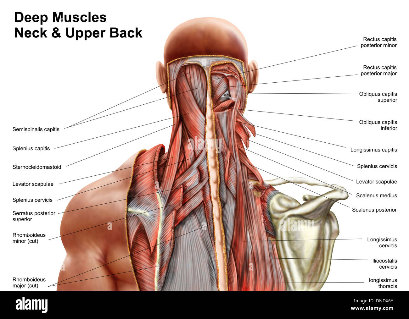
Diagram Of Nerves In Neck. Obliques Capitis Inferior Assist with head neck rotation. The spinal cord is a thick nerve trunk that forms the brains most important connection to the body and carries all signals to and from the brain that are not provided by the cranial nerves. The ansa cervicalis handle of the neck in Latin is a loop of nerves that lies superficial to the internal jugular vein composed of the C1 to C3 nerves. Nerves in the neck medically referred to as the cervical spine help transmit information along the pathways of the central and peripheral nervous system including sensory and motor skills processesthe cervical spine.
 Pin On Whats My Pain From pinterest.com
The musculocutaneous nerve travels along the front of the humerus and provides function to the Coracobrachialis biceps and brachialis muscles. Pharynx larynx glands common carotid internal carotid and external carotid arteries internal jugular vein facial glossopharyngeal vagus hypoglossal nerves. This is an article covering the formation branches course and distribution of the cervical plexus. Major nerves of the head and neck. An X-ray is one type of imaging tests that can aid in the diagnosis of a pinched nerve in the neck. The cervical spine is the top part of the spine.
Diagram of head and neck.
The ansa cervicalis handle of the neck in Latin is a loop of nerves that lies superficial to the internal jugular vein composed of the C1 to C3 nerves. Nerves Muscles Of The Head Neck Muscle Anatomy Human Anatomy Picture Muscle Anatomy. Obliques Capitis Inferior Assist with head neck rotation. Diagram Of The Neck Muscle. More specifically one end of the loop the superior root is derived from C1 and possibly C2 depending on the literature while the other the inferior root comes from C2 and C3. The branches of the trigeminal nerve V are represented in three different diagrams The ophthalmic nerve V1 in the orbital cavity with its main branches frontal nerve lacrimal nerve anterior and posterior ethmoidal nerve nasociliary nerve branch communicating with the ciliary ganglion supraorbital nerve supratrochlear nerve infra-trochlear nerve long ciliary nerves.
 Source: pinterest.com
C1 C2 and C3 the first three cervical nerves help control the head and neck including movements forward backward and to the sides. This is an article covering the formation branches course and distribution of the cervical plexus. Cervical Part of the Symphathetic Trunk. Rectus Capitis Lateralis Allows the neck to flex from side to side. Major nerves of the head and neck.
 Source: pinterest.com
The brachial plexus travels under the clavicle and through the armpit axilla. They are c1 through c8. The vessels transport a fluid. READ Bf3 Molecular orbital Diagram. You can click the image to magnify if you cannot see clearly.
 Source: pinterest.com
Nerves in the neck medically referred to as the cervical spine help transmit information along the pathways of the central and peripheral nervous system including sensory and motor skills processesthe cervical spine. Obliques Capitis Inferior Assist with head neck rotation. Superiorly - inferior border of mandible Medially - midline of neck Laterally - anterior border of sternocleidomastoid muscle Content. Diagram Of The Cutaneous Nerves Of The Head And Neck Stock Photo This diagram shows the veins present in the head and neck. The neck is unique in that it supports the weight of your head 10 to 11 pounds and allows a variety of headneck movement such as turning your head from side to side nodding and looking up.
 Source: pinterest.com
Major nerves supplying the Neck are. Anatomical diagrams of the spine and back imaios anatomical diagrams of the spine and back chest and lower back and the rotator muscles of the neck cranial nerves diagrams. The greater occipital nerve GON the lesser or small occipital nerve LON and the third or least. This image added by admin. C1 C2 and C3 the first three cervical nerves help control the head and neck including movements forward backward and to the sides.
 Source: id.pinterest.com
The cervical spine your neck is a complex structure making up the first region of the spinal column starting immediately below the skull and ending at the first thoracic vertebra. The neck is unique in that it supports the weight of your head 10 to 11 pounds and allows a variety of headneck movement such as turning your head from side to side nodding and looking up. These nerves can get pinchedcompressed due to injuries or underlying medical conditions. Cervical Part of the Symphathetic Trunk. The sensory cranial nerves are involved with the senses search as sight smell hearing and touch.
 Source: pinterest.com
The sensory cranial nerves are involved with the senses search as sight smell hearing and touch. Cutaneous Nerves of Neck. Superiorly - inferior border of mandible Medially - midline of neck Laterally - anterior border of sternocleidomastoid muscle Content. The cervical spine consists of eight different sets of nerves. The occipital nerves are a group of nerves that arise from the C2 and C3 spinal nerves12 They innervate the posterior scalp up as far as the vertex and other structures as well such as the ear2 There are three major occipital nerves in the human body.
 Source: pinterest.com
Obliques Capitis Superior Allows the neck to extend and flex to the side. An X-ray is one type of imaging tests that can aid in the diagnosis of a pinched nerve in the neck. Nerves in the neck medically referred to as the cervical spine help transmit information along the pathways of the central and peripheral nervous system including sensory and motor skills processes. You can click the image to magnify if you cannot see clearly. Nerves Muscles Of The Head Neck Muscle Anatomy Human Anatomy Picture Muscle Anatomy.
 Source: in.pinterest.com
Rectus Capitis Lateralis Allows the neck to flex from side to side. The muscles of the neck anatomical chart shows in beautiful detail the many anterior posterior inferior and lateral views of every muscle that makes up. Wandering through the neck and torso the vagus nerve communicates vital information from the brain to the heart and intestines. Rectus Capitis Lateralis Allows the neck to flex from side to side. The muscle anatomy of the head and neck is a fascinating area with the the neck also containing the 7 vertebrae of the part of the spine called the cervical curve.
 Source: pinterest.com
Whereas the motor nerves are responsible for controlling the movements and functions of muscles and glands cranial nerves supply sensory and motor information to areas of the head and neck. The muscle anatomy of the head and neck is a fascinating area with the the neck also containing the 7 vertebrae of the part of the spine called the cervical curve. Obliques Capitis Inferior Assist with head neck rotation. Cervical Part of the Symphathetic Trunk. The cervical spine your neck is a complex structure making up the first region of the spinal column starting immediately below the skull and ending at the first thoracic vertebra.
 Source: pinterest.com
The neck is unique in that it supports the weight of your head 10 to 11 pounds and allows a variety of headneck movement such as turning your head from side to side nodding and looking up. The brachial plexus is a group of nerves that branches from the neck cervical spine. The skin on the rear of the neck is supplied segmentally by cutaneous nerves originated from dorsal rami of C2 C3 and C4 spinal nerves. 1 The C2 dermatome handles sensation for the upper part of the head and the C3 dermatome covers the side of the face and back of the head. Cutaneous Nerves of Neck.
 Source: pinterest.com
This image added by admin. The occipital nerves are a group of nerves that arise from the C2 and C3 spinal nerves12 They innervate the posterior scalp up as far as the vertex and other structures as well such as the ear2 There are three major occipital nerves in the human body. An X-ray is one type of imaging tests that can aid in the diagnosis of a pinched nerve in the neck. Lower Back Muscles Diagram Unique Back Nerves Diagram Best Nerves the Abdomen Lower Back and. This is an article covering the formation branches course and distribution of the cervical plexus.
 Source: pinterest.com
Major nerves supplying the Neck are. The brachial plexus travels under the clavicle and through the armpit axilla. The cervical plexus is a network of nerves which forms from the anterior rami of C1-C4 within the prevertebral fascia in the posterior triangle of the neckIts branches can loosely be described as sensory of motor components. 2 C1 does not have a dermatome See The C1-C2 Vertebrae and Spinal Segment. Central nerve is a nerve cell that stays within the spinal cord or in the brain whereas the peripheral nerves are outside of the brain.
 Source: pinterest.com
Ventral Root of Spinal Nerves. Diagram of head and neck. The cervical spine consists of eight different sets of nerves. Central nerve is a nerve cell that stays within the spinal cord or in the brain whereas the peripheral nerves are outside of the brain. For example the nodes in the neck are called cervical nodes after the cervical part of the vertebral column and mandibular nodes after the mandible or jawbone.
 Source: pinterest.com
Levator Scapulae Responsible for movement of the scapula shoulder blade in an upward and downward motion. More specifically one end of the loop the superior root is derived from C1 and possibly C2 depending on the literature while the other the inferior root comes from C2 and C3. The greater occipital nerve GON the lesser or small occipital nerve LON and the third or least. Anatomical diagrams of the spine and back imaios anatomical diagrams of the spine and back chest and lower back and the rotator muscles of the neck cranial nerves diagrams. Nicole long a diagram showing nerves in the head and neck.
 Source: pinterest.com
An X-ray is one type of imaging tests that can aid in the diagnosis of a pinched nerve in the neck. Nerves Muscles Of The Head Neck Muscle Anatomy Human Anatomy Picture Muscle Anatomy. Major nerves supplying the Neck are. The skin on the rear of the neck is supplied segmentally by cutaneous nerves originated from dorsal rami of C2 C3 and C4 spinal nerves. The branches of the trigeminal nerve V are represented in three different diagrams The ophthalmic nerve V1 in the orbital cavity with its main branches frontal nerve lacrimal nerve anterior and posterior ethmoidal nerve nasociliary nerve branch communicating with the ciliary ganglion supraorbital nerve supratrochlear nerve infra-trochlear nerve long ciliary nerves.
 Source: pinterest.com
We think this is the most useful anatomy picture that you need. Lower Back Muscles Diagram Unique Back Nerves Diagram Best Nerves the Abdomen Lower Back and. Superiorly - inferior border of mandible Medially - midline of neck Laterally - anterior border of sternocleidomastoid muscle Content. Major nerves supplying the Neck are. Cutaneous Nerves of Neck.
 Source: pinterest.com
This is an article covering the formation branches course and distribution of the cervical plexus. The vessels transport a fluid. Rectus Capitis Lateralis Allows the neck to flex from side to side. Wandering through the neck and torso the vagus nerve communicates vital information from the brain to the heart and intestines. The greater occipital nerve GON the lesser or small occipital nerve LON and the third or least.
 Source: pinterest.com
Nerves Muscles Of The Head Neck Muscle Anatomy Human Anatomy Picture Muscle Anatomy. The cervical spine is the top part of the spine. Anatomical diagrams of the spine and back imaios anatomical diagrams of the spine and back chest and lower back and the rotator muscles of the neck cranial nerves diagrams. The cervical spine your neck is a complex structure making up the first region of the spinal column starting immediately below the skull and ending at the first thoracic vertebra. The muscle anatomy of the head and neck is a fascinating area with the the neck also containing the 7 vertebrae of the part of the spine called the cervical curve.
This site is an open community for users to submit their favorite wallpapers on the internet, all images or pictures in this website are for personal wallpaper use only, it is stricly prohibited to use this wallpaper for commercial purposes, if you are the author and find this image is shared without your permission, please kindly raise a DMCA report to Us.
If you find this site serviceableness, please support us by sharing this posts to your favorite social media accounts like Facebook, Instagram and so on or you can also save this blog page with the title diagram of nerves in neck by using Ctrl + D for devices a laptop with a Windows operating system or Command + D for laptops with an Apple operating system. If you use a smartphone, you can also use the drawer menu of the browser you are using. Whether it’s a Windows, Mac, iOS or Android operating system, you will still be able to bookmark this website.