Diagram of sinus cavity
Home » Background » Diagram of sinus cavityYour Diagram of sinus cavity images are ready in this website. Diagram of sinus cavity are a topic that is being searched for and liked by netizens now. You can Get the Diagram of sinus cavity files here. Download all royalty-free photos and vectors.
If you’re searching for diagram of sinus cavity images information related to the diagram of sinus cavity topic, you have come to the right blog. Our site always gives you hints for viewing the highest quality video and image content, please kindly search and locate more informative video content and graphics that fit your interests.
Diagram Of Sinus Cavity. Interactive diagrams show sinus cavity locations and help visualize sinusitis the most common type of sinus infection. There are two types of sinusitis. The paranasal sinuses surround and drain into the nasal cavity. There are several sets of sinuses in the head.
 Image Result For Cavernous Sinus Anatomia Del Cerebro Humano Aparatos De Ortodoncia Anatomia Del Ojo From pinterest.com
- nasal cavity diagram stock illustrations. The cavernous sinus is not only involved in the venous drainage of the brain but also facilitates the passage of numerous vessels and cranial nerves. The maxillary sinuses are behind the cheeks. Sinuses are hollow air-filled cavities that are located throughout the skull. The largest sinus cavities are about an inch across. Heres a pretty good diagram of the anatomy although for some reason this cartoonIn general a neti pot is an excellent tool to perform nasal irrigation and reduce general sinus congestion.
Maxillary sinuses the largest cavities these two sit behind the cheekbones.
They are divided into front middle and. Your Kidneys How. The ethmoid sinuses lie under the inside corners of the eyes. Neck Strain Causes And Rem. Acute sinusitis - an infection that is often triggered by the flu or cold. Sinuses in back head diagram anatomy organ the sinuses in back head diagram can be your consideration when thinking of about anatomy diagram when showing this sinuses in back head diagram i can guarantee to aspire you for this time we gather some pictures of sinuses in back head diagram and each of them displaying some new ideas sinus cavities in the head.
 Source: pinterest.com
The ethmoid sinuses are not single sacs. 40 Sinus Cavity Diagram Nasal Sinus Cavities Me. The flu or cold virus attacks your sinuses causing them to swell and become narrow. This might be helpful for example to understand a doctors diagnosis or your X-ray. Your Kidneys How.
 Source: pinterest.com
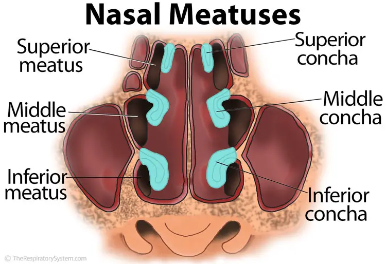
A sinus is a sac or cavity in any organ or tissue or an abnormal cavity or passage caused by the destruction of tissue. The sphenoid sinuses are located behind the ethmoid sinuses. The anterior ethmoidal sinuses as well as the frontal and maxillary sinuses drain into the middle meatus the opening marked with a crescent groove called the semilunar hiatus on the lateral wall of the cavity 19. Acute sinusitis - an infection that is often triggered by the flu or cold. Each sinus is lined by a ciliated pseudostratified.
 Source: pinterest.com
32 Heavy Cartoon Drawing Png - Self Plant Drawing. The sinuses are a connected system of hollow cavities in the skull. Aug 09 2011 sinus cavities diagram published on aug 9 2011 fundamentally situated in the skull human sinus cavities are the passageways mainly found in the areas around the face. The middle ethmoidal sinuses form a bump the. The dorsal body cavity protects organs of the nervous system and has two subdivisions.
 Source: pinterest.com
Vintage anatomy print of the human leg showcasing the veins and arteries. Sinuses In Back Of Head Diagram. 14 Lorax Cartoon Drawing - The Lorax Coloring Pag. Each cavity is the continuation of one of the two nostrils. The sphenoid sinuses are located in the sphenoid bone near the optic nerve and the pituitary gland on the side of the skull.
 Source: pinterest.com
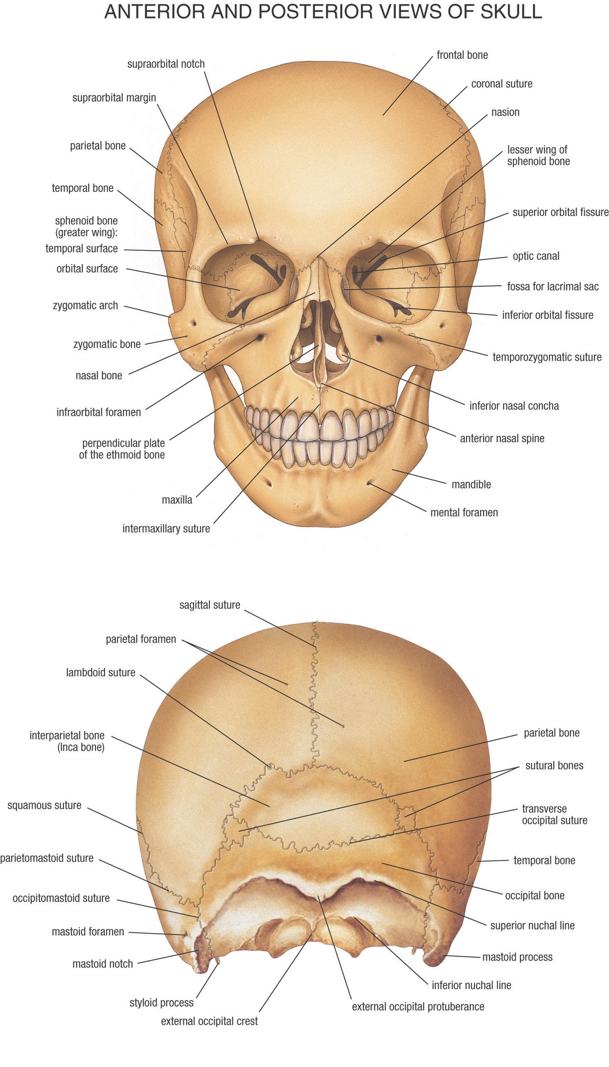
Sinusitis is caused by an inflammation of your sinus cavities that causes redness swelling mucus and pain. The maxillary sinuses are behind the cheeks. The ethmoid sinuses are. Interactive diagrams show sinus cavity locations and help visualize sinusitis the most common type of sinus. A multi view of the human skull showing the bone breakdown.
 Source: pinterest.com
The anterior ethmoidal sinuses as well as the frontal and maxillary sinuses drain into the middle meatus the opening marked with a crescent groove called the semilunar hiatus on the lateral wall of the cavity 19. Sinuses in back head diagram anatomy organ the sinuses in back head diagram can be your consideration when thinking of about anatomy diagram when showing this sinuses in back head diagram i can guarantee to aspire you for this time we gather some pictures of sinuses in back head diagram and each of them displaying some new ideas sinus cavities in the head. We also go over sinusitis signs and care. Heres a pretty good diagram of the anatomy although for some reason this cartoonIn general a neti pot is an excellent tool to perform nasal irrigation and reduce general sinus congestion. Whatever the cause neti pots are best known to help get the inside of your nose in a healthier less irritated state by flushing out any unwanted invaders and excess mucus.
 Source: pinterest.com
Aug 09 2011 sinus cavities diagram published on aug 9 2011 fundamentally situated in the skull human sinus cavities are the passageways mainly found in the areas around the face. They are a collection of six to 12 small air cells that open independently into the nasal cavity. The paranasal sinuses are air-filled extensions of the nasal cavity. The middle ethmoidal sinuses form a bump the. Maxillary sinuses the largest cavities these two sit behind the cheekbones.
 Source: pinterest.com
The medullary cavity contains red bone marrow during childhood eventually turning into yellow bone marrow after puberty. The ethmoid sinuses lie under the inside corners of the eyes. Acute sinusitis - an infection that is often triggered by the flu or cold. Human body organ diagram front view. Others are much smaller.
 Source: pinterest.com
Human body organ diagram front view. We also go over sinusitis signs and care. Sinuses In Back Of Head Diagram. These are absent at birth and in approximately 5 of the adult population. Whatever the cause neti pots are best known to help get the inside of your nose in a healthier less irritated state by flushing out any unwanted invaders and excess mucus.
 Source: pinterest.com
The cavernous sinus is not only involved in the venous drainage of the brain but also facilitates the passage of numerous vessels and cranial nerves. Where are sinus cavities located sinus cavity diagram this is your largest sinus cavity see the diagram to an idea ethmoid sinus cavity the ethmoid sinus cavity is located right in between your eyes right over your nasal bridge more specifically sphenoid sinus cavity the sphenoid sinus cavity a quite odd name indeed is located in the bones behind the nasal cavity see the diagram for a better idea of where it is. Your Kidneys How. Each sinus is lined by a ciliated pseudostratified. They are a collection of six to 12 small air cells that open independently into the nasal cavity.
 Source: pinterest.com
- nasal cavity diagram stock illustrations. The maxillary sinuses are behind the cheeks. The paranasal sinuses surround and drain into the nasal cavity. This sinus is located in the maxilla bone under the eye contains three recesses and is shaped like a pyramid. 40 Sinus Cavity Diagram Nasal Sinus Cavities Me.
 Source: pinterest.com
The anterior ethmoidal sinuses as well as the frontal and maxillary sinuses drain into the middle meatus the opening marked with a crescent groove called the semilunar hiatus on the lateral wall of the cavity 19. Interactive diagrams show sinus cavity locations and help visualize sinusitis the most common type of sinus infection. Vintage anatomy print features the hand of a human skeleton with bones labeled. The anterior ethmoidal sinuses as well as the frontal and maxillary sinuses drain into the middle meatus the opening marked with a crescent groove called the semilunar hiatus on the lateral wall of the cavity 19. - nasal cavity diagram stock illustrations.
This site is an open community for users to do sharing their favorite wallpapers on the internet, all images or pictures in this website are for personal wallpaper use only, it is stricly prohibited to use this wallpaper for commercial purposes, if you are the author and find this image is shared without your permission, please kindly raise a DMCA report to Us.
If you find this site good, please support us by sharing this posts to your favorite social media accounts like Facebook, Instagram and so on or you can also bookmark this blog page with the title diagram of sinus cavity by using Ctrl + D for devices a laptop with a Windows operating system or Command + D for laptops with an Apple operating system. If you use a smartphone, you can also use the drawer menu of the browser you are using. Whether it’s a Windows, Mac, iOS or Android operating system, you will still be able to bookmark this website.