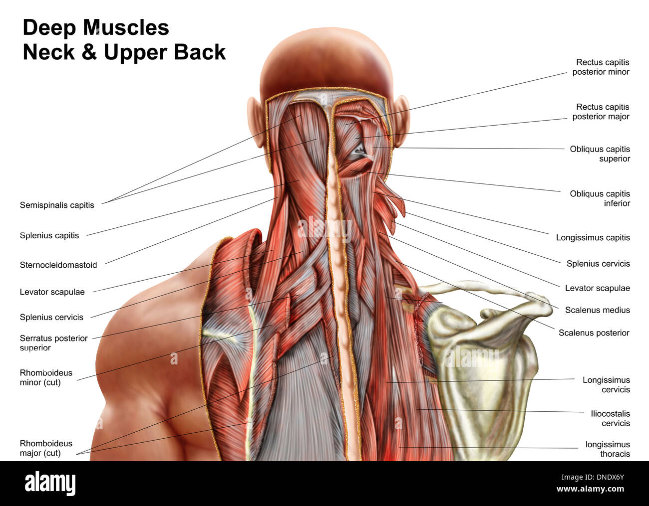
Diagram of the back of the head
Home » Wallpapers » Diagram of the back of the headYour Diagram of the back of the head images are available in this site. Diagram of the back of the head are a topic that is being searched for and liked by netizens now. You can Get the Diagram of the back of the head files here. Get all royalty-free photos and vectors.
If you’re searching for diagram of the back of the head pictures information linked to the diagram of the back of the head keyword, you have come to the ideal blog. Our site frequently provides you with suggestions for viewing the maximum quality video and image content, please kindly hunt and locate more enlightening video content and graphics that match your interests.
Diagram Of The Back Of The Head. The post auricular or mastoid nodes are located behind the ears and drain the back of the neck part of. The head or headstock is where you tune the guitar. Given below is a. These are easiest to see on the side of the head where the temporal bone meets the parietal bone and in the back where the occipital bone adjoins the.
 The Human Muscle System Neck Muscle Anatomy Muscles Of The Neck Muscle Anatomy From pinterest.com
Finally the image is then projected in the cortex. An anatomical diagram consisting of a vertical cross-section of the human head and showing the relations of the nasal and buccal cavities and the. Other muscles are small and cover much less space. May 11 2018 - Muscle and anatomy are two words that are often heard when you are studying science. The backs muscles start at the top of the back named the cervical vertebrae and go to the tailbone also named the coccyx. These two regions are responsible for most of the movement in the back allowing you to bend and twist.
The Mayo Clinic says that these factors can be chemical activity in your brain the nerves of blood vessels surrounding your skull or the muscles at the back of the head and neck.
The head or headstock is where you tune the guitar. Choose from a nice collection of body outline front and back worksheets. An anatomical diagram consisting of a vertical cross-section of the human head and showing the relations of the nasal and buccal cavities and the. These two regions are responsible for most of the movement in the back allowing you to bend and twist. The back muscles represented on an anatomical chart and on a schematic view of the origin and insertion of the proper muscles of the back iliocostal muscle of the neck lumbar lumbar and thoracic parts longissimus muscles of head neck and thorax the spinalis muscles of the neck and thorax semispinalis muscle of the head neck and thorax lateral. Shoulder skeleton diagram with head and deltoid tubercle of humerus scapula skeletal structure anatomy of neck and shoulder stock illustrations Lower back pain Lower back pain anatomy of neck and shoulder stock pictures royalty-free photos images.
 Source: sk.pinterest.com
These two regions are responsible for most of the movement in the back allowing you to bend and twist. The thoracic spine allows little movement and therefore is injured less often. If someone wants a healthy and good life one must understand his body. See human head diagram stock video clips. Working individually these muscles rotate the head or flex the neck laterally to the left or right.
 Source: pinterest.com
These run from the transverse processes to the neck and tubercle of the ribs respectively. The backs muscles start at the top of the back named the cervical vertebrae and go to the tailbone also named the coccyx. A broad sheet of muscle fibers extending from the collarbone to the angle of the jaw. The separation of the cranial bone plates at time of birth facilitate passage of the head of the fetus through the mothers birth canal or pelvic girdle. Lymph Nodes of the Neck.
 Source: pinterest.com
Costovertebral joints and ligaments. The outer portion contains neurons and the inner area communicates with the cerebral cortex. The separation of the cranial bone plates at time of birth facilitate passage of the head of the fetus through the mothers birth canal or pelvic girdle. An anatomical diagram consisting of a vertical cross-section of the human head and showing the relations of the nasal and buccal cavities and the. The cerebellum little brain is a fist-sized portion of the brain located at the back of the head below the temporal and occipital lobes and above the brainstem.
 Source: pinterest.com
The human body consists of many muscles. - human head diagram stock illustrations. Shoulder skeleton diagram with head and deltoid tubercle of humerus scapula skeletal structure anatomy of neck and shoulder stock illustrations Lower back pain Lower back pain anatomy of neck and shoulder stock pictures royalty-free photos images. Choose from a nice collection of body outline front and back worksheets. The human body consists of many muscles.
 Source: pinterest.com
The back muscles represented on an anatomical chart and on a schematic view of the origin and insertion of the proper muscles of the back iliocostal muscle of the neck lumbar lumbar and thoracic parts longissimus muscles of head neck and thorax the spinalis muscles of the neck and thorax semispinalis muscle of the head neck and thorax lateral. Costovertebral joints and ligaments. Cervical Spine Diagram Stress in the spine is greatest in the cervical neck and lumbar lower back areas. The outer portion contains neurons and the inner area communicates with the cerebral cortex. The parietal bones and occipital bone can overlap each other in the birth canal.
 Source: pinterest.com
The head or headstock is where you tune the guitar. They move the head in every direction pulling the skull and jaw towards the shoulders spine and scapula. The backs muscles start at the top of the back named the cervical vertebrae and go to the tailbone also named the coccyx. Certain back muscles extend to other areas like the shoulders upper arms and thighs. The neck is where you hold the guitar in your left hand if youre right handed or your right hand if youre left handed.
 Source: pinterest.com
Certain back muscles extend to other areas like the shoulders upper arms and thighs. In a newborn the junction of the parietal bones with the frontal and occipital bones form the anterior front and posterior back fontanelle or soft spots. The cerebellum little brain is a fist-sized portion of the brain located at the back of the head below the temporal and occipital lobes and above the brainstem. 11693 human head diagram stock photos vectors and illustrations are available royalty-free. The outer portion contains neurons and the inner area communicates with the cerebral cortex.
 Source: pinterest.com
11693 human head diagram stock photos vectors and illustrations are available royalty-free. See human head diagram stock video clips. The outer portion contains neurons and the inner area communicates with the cerebral cortex. In a newborn the junction of the parietal bones with the frontal and occipital bones form the anterior front and posterior back fontanelle or soft spots. Certain back muscles extend to other areas like the shoulders upper arms and thighs.
 Source: pinterest.com
The Mayo Clinic says that these factors can be chemical activity in your brain the nerves of blood vessels surrounding your skull or the muscles at the back of the head and neck. It is opposite from the chest and the vertebral column runs down the back. Each visual cortex receives raw sensory information from the outside half of the eye present on the same side and from the inside half of the eye present on the other side of the head. Choose from a nice collection of body outline front and back worksheets. Some of these muscles are quite large and cover broad areas.
 Source: pinterest.com
Certain back muscles extend to other areas like the shoulders upper arms and thighs. Guitar Parts Diagram - Main Parts Of The Guitar. These two regions are responsible for most of the movement in the back allowing you to bend and twist. The CVJ is one of the unique and complex areas of your body as this is where your brain transitions to your spine. It comprises the vertebral column spine and two compartments of back muscles.
 Source: pinterest.com
The backs muscles start at the top of the back named the cervical vertebrae and go to the tailbone also named the coccyx. Side view of a males brain showcasing the venous system thalamus and cerebellum. Some of these muscles are quite large and cover broad areas. The CVJ is one of the unique and complex areas of your body as this is where your brain transitions to your spine. May 11 2018 - Muscle and anatomy are two words that are often heard when you are studying science.
 Source: pinterest.com
They move the head in every direction pulling the skull and jaw towards the shoulders spine and scapula. In a newborn the junction of the parietal bones with the frontal and occipital bones form the anterior front and posterior back fontanelle or soft spots. The back functions are many such as to house and protect the spinal cord hold the body and head upright and adjust the movements of the upper and lower limbs. Some of these muscles are quite large and cover broad areas. These are easiest to see on the side of the head where the temporal bone meets the parietal bone and in the back where the occipital bone adjoins the.
 Source: pinterest.com
As illustrated in the diagram below the guitar like humans has a head neck and body. Side view of a males brain showcasing the venous system thalamus and cerebellum. These two regions are responsible for most of the movement in the back allowing you to bend and twist. The back functions are many such as to house and protect the spinal cord hold the body and head upright and adjust the movements of the upper and lower limbs. 1 Also some people are more prone to headaches than others.
 Source: pinterest.com
The parietal bones and occipital bone can overlap each other in the birth canal. Like the cerebral cortex it has two hemispheres. - human head diagram stock illustrations. 11693 human head diagram stock photos vectors and illustrations are available royalty-free. The CVJ is one of the unique and complex areas of your body as this is where your brain transitions to your spine.
 Source: pinterest.com
Choose from a nice collection of body outline front and back worksheets. Other muscles are small and cover much less space. The neck is where you hold the guitar in your left hand if youre right handed or your right hand if youre left handed. The head or headstock is where you tune the guitar. Human body outlines are available for PDF format.
 Source: id.pinterest.com
These run from the transverse processes to the neck and tubercle of the ribs respectively. It is opposite from the chest and the vertebral column runs down the back. Each of a pair of long muscles that connect the sternum clavicle and mastoid process of the temporal bone and serve to turn and nod the head. The back muscles represented on an anatomical chart and on a schematic view of the origin and insertion of the proper muscles of the back iliocostal muscle of the neck lumbar lumbar and thoracic parts longissimus muscles of head neck and thorax the spinalis muscles of the neck and thorax semispinalis muscle of the head neck and thorax lateral. An anatomical diagram consisting of a vertical cross-section of the human head and showing the relations of the nasal and buccal cavities and the.
 Source: pinterest.com
Shoulder skeleton diagram with head and deltoid tubercle of humerus scapula skeletal structure anatomy of neck and shoulder stock illustrations Lower back pain Lower back pain anatomy of neck and shoulder stock pictures royalty-free photos images. Shoulder skeleton diagram with head and deltoid tubercle of humerus scapula skeletal structure anatomy of neck and shoulder stock illustrations Lower back pain Lower back pain anatomy of neck and shoulder stock pictures royalty-free photos images. The parietal bones and occipital bone can overlap each other in the birth canal. Cervical Spine Diagram Stress in the spine is greatest in the cervical neck and lumbar lower back areas. It is opposite from the chest and the vertebral column runs down the back.
 Source: pinterest.com
- human head diagram stock illustrations. In a newborn the junction of the parietal bones with the frontal and occipital bones form the anterior front and posterior back fontanelle or soft spots. A broad sheet of muscle fibers extending from the collarbone to the angle of the jaw. Human face muscles old anatomy drawings cerebral hemispheres vintage anatomy head diagram the brain human nervous system chart human brain anatomy diagram hemispheres of the brain head diagram old. The back is the body region between the neck and the gluteal regions.
This site is an open community for users to do sharing their favorite wallpapers on the internet, all images or pictures in this website are for personal wallpaper use only, it is stricly prohibited to use this wallpaper for commercial purposes, if you are the author and find this image is shared without your permission, please kindly raise a DMCA report to Us.
If you find this site helpful, please support us by sharing this posts to your own social media accounts like Facebook, Instagram and so on or you can also save this blog page with the title diagram of the back of the head by using Ctrl + D for devices a laptop with a Windows operating system or Command + D for laptops with an Apple operating system. If you use a smartphone, you can also use the drawer menu of the browser you are using. Whether it’s a Windows, Mac, iOS or Android operating system, you will still be able to bookmark this website.