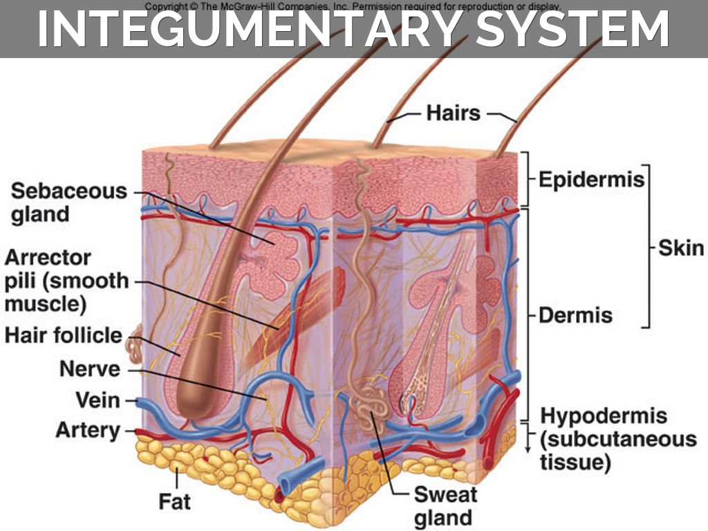
Diagram of the dermis
Home » Wallpapers » Diagram of the dermisYour Diagram of the dermis images are available. Diagram of the dermis are a topic that is being searched for and liked by netizens now. You can Find and Download the Diagram of the dermis files here. Find and Download all royalty-free photos.
If you’re looking for diagram of the dermis images information connected with to the diagram of the dermis topic, you have visit the ideal site. Our website frequently gives you suggestions for viewing the maximum quality video and picture content, please kindly search and locate more informative video content and images that fit your interests.
Diagram Of The Dermis. The integumentary system skin functions of the integumentary system the epidermis thin outer layer of skin the dermis thick. They also contain anchoring fibrils. Lymphatic capillaries and vessels. An example is the skin on the.
 Human Skin Cells Labeled Google Search Subcutaneous Tissue Skin Structure Epidermis From pinterest.com
We hope this picture Epidermis Dermis Anatomical Location Diagram can help you study and research. The dermis is a tough and elastic layer containing white fibrous tissue interlaced with yellow elastic fibers. This layer of the dermis projects into the stratum basale of the epidermis and forms finger-like dermal papillae. The role of the dermis is to support and protect the skin and deeper layers assist in thermoregulation and aid in sensation. The collagen and elastin fibres are loosely arranged in the layer. It is the superficial layer of the dermis.
The ducts can also open directly on the surface of the skin as seen on the lips and buccal mucosa.
They are also involved in regulating body temperature. An example is the skin on the. While the epidermis covers your body. Humans shed around 500 million skin cells each day. The outermost layer of the skin is. It is the layer that holds all the blood vessels nerves hair follicles collagen and sweat glands.
 Source: pinterest.com
It is the superficial layer of the dermis. The epidermis dermis and hypodermisThere are also sweat glands and hairs which have sebaceous glands and a smooth muscle called the arrector pili muscle associated with them. An example is the skin on the. Hair follicle microscope layers of dermis skin istology structure of dermis structure skin the skin anatomy the structure of the skin subcutaneous layer the skin an layersskin diagrams. However the fine collagen network of interwoven.
 Source: pinterest.com
The integumentary system skin functions of the integumentary system the epidermis thin outer layer of skin the dermis thick. 18507 dermis stock photos vectors and illustrations are available royalty-free. Undoubtedly the skin is the largest organ in the human body. The dermis is a tough and elastic layer containing white fibrous tissue interlaced with yellow elastic fibers. There are three main layers.
 Source: pinterest.com
However the fine collagen network of interwoven. Outer papillary layer deep reticular layer Characteristics of Dermis Dense Irregular Connective Tissue. Practice labeling the layers of the skin. DERMIS What are the structures and functions of the dermis. As you can see in the skin diagram many structures are embedded in the dermis including.
 Source: pinterest.com
Tips and advice on skin. While the epidermis covers your body. The dermis the epidermis. Labeling The Integumentary System Diagram of the skin -. This layer of the dermis projects into the stratum basale of the epidermis and forms finger-like dermal papillae.
 Source: pinterest.com
Epidermis dermis and hypodermis which contain certain sublayers. Sweat glands and their ducts. Normal Skin Anatomy Diagram Skin 1 The Structure And Functions Of The Skin Nursing Times -. The dermis the epidermis. Which is the thickest layer.
 Source: pinterest.com
The dermis is tucked away between the epidermis and hypodermis. The dermis is the layer of the skin present beneath the epidermis of the skin. This papillary layer can protrude into the epidermis and giving rise to dermal papillae. An example is the skin on the. Sebaceous glands are small saccular structures located in the dermis which cover most of the body.
 Source: pinterest.com
The epidermis dermis and hypodermis. Learn vocabulary terms and more with flashcards games and other study tools. As you can see in the skin diagram many structures are embedded in the dermis including. The Dermis Is located between epidermis and subcutaneous layer Anchors epidermal accessory structures hair follicles sweat glands Has 2 layers. Lymphatic capillaries and vessels.
 Source: pinterest.com
DERMIS What are the structures and functions of the dermis. This papillary layer can protrude into the epidermis and giving rise to dermal papillae. First the density of dermal papillae is higher compared with healthy skin. This stained slide shows the two components of the dermisthe papillary layer and the reticular layer. The dermis the epidermis.
 Source: pinterest.com
They also contain anchoring fibrils. The epidermis dermis and hypodermisThere are also sweat glands and hairs which have sebaceous glands and a smooth muscle called the arrector pili muscle associated with them. Full color diagram of skin anatomy. As you can see in the skin diagram many structures are embedded in the dermis including. They consist of a cluster of secretory acini which is continued by a duct which opens into the dermal pilary canal of the hair follicle.
 Source: pinterest.com
Learn vocabulary terms and more with flashcards games and other study tools. 18507 dermis stock photos vectors and illustrations are available royalty-free. First the density of dermal papillae is higher compared with healthy skin. See dermis stock video clips. Dermis or corium layer.
 Source: pinterest.com
First the density of dermal papillae is higher compared with healthy skin. Normal Skin Anatomy Diagram Skin 1 The Structure And Functions Of The Skin Nursing Times -. First the density of dermal papillae is higher compared with healthy skin. We hope this picture Epidermis Dermis Anatomical Location Diagram can help you study and research. Outer papillary layer deep reticular layer Characteristics of Dermis Dense Irregular Connective Tissue.
 Source: pinterest.com
An example is the skin on the. Sweat glands and their ducts. Find normal skin anatomy stock images in hd and millions of other. An example is the skin on the. Learn vocabulary terms and more with flashcards games and other study tools.
 Source: pinterest.com
This diagram shows the layers found in skin. An example is the skin on the. They also contain anchoring fibrils. The epidermis dermis and hypodermis. Normal Skin Anatomy Diagram Skin 1 The Structure And Functions Of The Skin Nursing Times -.
 Source: pinterest.com
Outer papillary layer deep reticular layer Characteristics of Dermis Dense Irregular Connective Tissue. The role of the dermis is to support and protect the skin and deeper layers assist in thermoregulation and aid in sensation. Tips and advice on skin. The ducts can also open directly on the surface of the skin as seen on the lips and buccal mucosa. The presence of papillae is still evident when moving deeper into the tissue where the space around them starts to be filled with collagen.
 Source: pinterest.com
The integumentary system skin functions of the integumentary system the epidermis thin outer layer of skin the dermis thick. As you can see in the skin diagram many structures are embedded in the dermis including. It is the layer that holds all the blood vessels nerves hair follicles collagen and sweat glands. For more anatomy content please follow us and visit our website. First the density of dermal papillae is higher compared with healthy skin.
 Source: pinterest.com
This stained slide shows the two components of the dermisthe papillary layer and the reticular layer. An example is the skin on the. Lymphatic capillaries and vessels. There are three main layers. While the epidermis covers your body.
 Source: pinterest.com
The subcutaneous layer under the dermis is made up of connective tissue and fat a good insulator. An example is the skin on the. Both are made of connective tissue with fibers of collagen extending from one to the other making the. The epidermis dermis and hypodermisThere are also sweat glands and hairs which have sebaceous glands and a smooth muscle called the arrector pili muscle associated with them. The subcutaneous layer under the dermis is made up of connective tissue and fat a good insulator.
 Source: pinterest.com
Full color diagram of skin anatomy. For more anatomy content please follow us and visit our website. Find normal skin anatomy stock images in hd and millions of other. At a depth of 170 μm from the skin surface dermis starts having a similar morphology with respect to healthy skin. Practice labeling the layers of the skin.
This site is an open community for users to do submittion their favorite wallpapers on the internet, all images or pictures in this website are for personal wallpaper use only, it is stricly prohibited to use this wallpaper for commercial purposes, if you are the author and find this image is shared without your permission, please kindly raise a DMCA report to Us.
If you find this site serviceableness, please support us by sharing this posts to your preference social media accounts like Facebook, Instagram and so on or you can also save this blog page with the title diagram of the dermis by using Ctrl + D for devices a laptop with a Windows operating system or Command + D for laptops with an Apple operating system. If you use a smartphone, you can also use the drawer menu of the browser you are using. Whether it’s a Windows, Mac, iOS or Android operating system, you will still be able to bookmark this website.