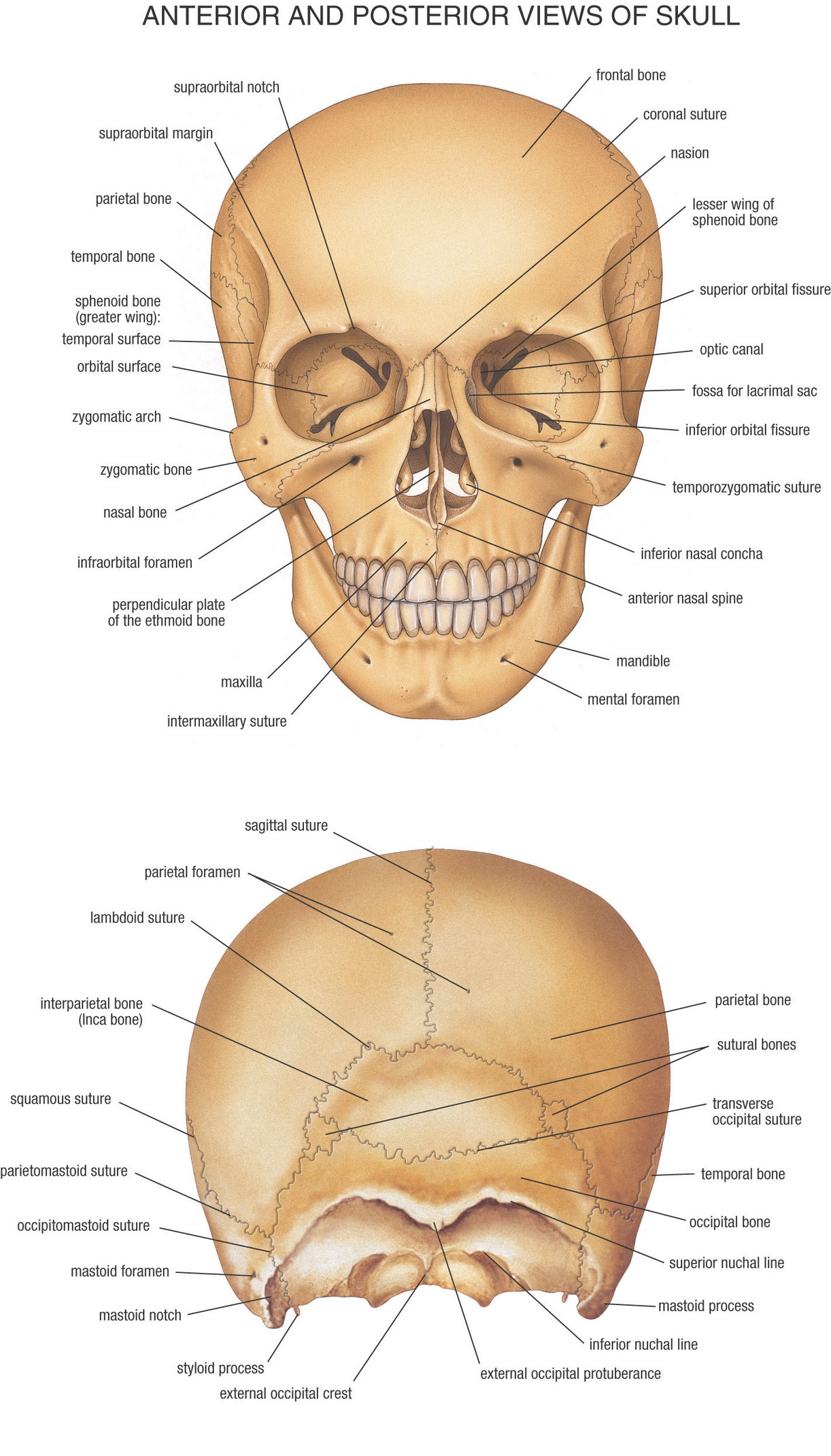
Diagram of the skull
Home » Wallpapers » Diagram of the skullYour Diagram of the skull images are available. Diagram of the skull are a topic that is being searched for and liked by netizens now. You can Get the Diagram of the skull files here. Download all free vectors.
If you’re looking for diagram of the skull images information related to the diagram of the skull keyword, you have come to the right blog. Our website frequently provides you with hints for viewing the maximum quality video and picture content, please kindly search and find more enlightening video articles and images that match your interests.
Diagram Of The Skull. The skull is a bony structure that supports the face and forms a protective cavity for the brain. Use this PowerPoint template to explain the anatomy of a skull and find out. The cranium skull is the skeletal structure of the head that supports the face and protects the brain. The facial bones underlie the facial structures form the nasal.
 The Skeletal System Anatomy Bones Human Anatomy And Physiology Anatomy From pinterest.com
As you can see in the above diagram there are around 24 labels and forgetting one of them is quite possible. Develop a good way to remember the cranial bone markings types definition and names including the frontal bone occipital bone parietal bone temporal bone sphenoid bone and ethmoid bone using this. The main side of the skull. Lets start by the first anterior part of skull diagram. It is subdivided into the facial bones and the brain case or cranial vault. The facial bones underlie the facial structures form the nasal.
Lets start by the first anterior part of skull diagram.
Weve created a blank skull diagram free for you to download as A PDF below. Facial BonesDental AnatomyMusculoskeletal SystemSkeletonsNursesSkull AnatomyAnatomy. It acts as a cover to the brain and supports the structure of the face. The two parts of the skull are the cranium and facial bones. It is subdivided into the facial bones and the brain case or cranial vault. As you can see in the above diagram there are around 24 labels and forgetting one of them is quite possible.
 Source: pinterest.com
The human diagram is labeled and you can learn each part of the body to understand the function and location. To be more precise the squamous part of the frontal squamous part of occipital bone above the superior nuchal line parietal bones squamous portion of the temporal bone and the great wing of sphenoid bone forms the calvaria. It acts as a cover to the brain and supports the structure of the face. The cranium skull is the skeletal structure of the head that supports the face and protects the brain. It is subdivided into the facial bones and the brain caseSkull anatomy unlabeled In this image you will find Skull anatomy in it.
 Source: pinterest.com
In the CT data the upper teeth and lower teeth were joined as one mesh therefore I completely remodelled some of the teeth so that the mandible could be detached from the cranium and can be. The cranium and mandible was exported from CT data. It is comprised of many bones formed by intramembranous ossification which are joined together by sutures fibrous joints. Firstly divide the skeleton into small groupsthe skull the. It acts as a cover to the brain and supports the structure of the face.
 Source: pinterest.com
Use this PowerPoint template to explain the anatomy of a skull and find out. Skull system anatomy of skull anatomy skull skull lateral view skull anatomy occipital medical skeleton named facial bone anatomy mandible maxilla human anatomy lateral skull anatomy. The human diagram is labeled and you can learn each part of the body to understand the function and location. The cranium skull is the skeletal structure of the head that supports the face and protects the brain. Skull Anatomy The Human Skull Laminated Anatomical Chart The Human Skull Anatomical Chart shows anterior and lateral aspects of the skull base of skull including inner surface sagittal section through skull horizontal section through maxilla mandible coronal section through anterior skull ethmoid bone sphenoid bone lateral wall of left nasal cavity and medial wall of.
 Source: sk.pinterest.com
These bones articulate through three sutures. The skull is a structure of bones that protects the brain and helps to support the features of the face. Skull Anatomy PowerPoint Diagram. These skull diagrams use different colors to show different parts and also labels to show a number of important skull parts. These printable anatomy diagrams are designed to guide you in studying the structure of the human skull.
 Source: pinterest.com
The occipital bone forms the base of the skull. The facial bones underlie the facial structures form the nasal cavity enclose the eyeballs and support the teeth of the upper and lower jaws. Between the frontal and parietal bones. The cranium skull is the skeletal structure of the head that supports the face and protects the brain. The skull of humans is said to be dicondylic.
 Source: pinterest.com
The skull is made up of 22 bones that articulate with each other - 8 cranial bones and 14 facial bones. You can also download the labeled version and use this to make some notes. The skull is a bony structure that supports the face and forms a protective cavity for the brain. Weve created a blank skull diagram free for you to download as A PDF below. Human Anatomy Diagram Of Skull With Radiographic Land Marks.
 Source: pinterest.com
These bones articulate through three sutures. Firstly divide the skeleton into small groupsthe skull the. Ready to test your knowledge. In the CT data the upper teeth and lower teeth were joined as one mesh therefore I completely remodelled some of the teeth so that the mandible could be detached from the cranium and can be. 115084 human skull anatomy stock photos vectors and illustrations are available royalty-free.
 Source: pinterest.com
Skull system anatomy of skull anatomy skull skull lateral view skull anatomy occipital medical skeleton named facial bone anatomy mandible maxilla human anatomy lateral skull anatomy. In the CT data the upper teeth and lower teeth were joined as one mesh therefore I completely remodelled some of the teeth so that the mandible could be detached from the cranium and can be. To be more precise the squamous part of the frontal squamous part of occipital bone above the superior nuchal line parietal bones squamous portion of the temporal bone and the great wing of sphenoid bone forms the calvaria. 115084 human skull anatomy stock photos vectors and illustrations are available royalty-free. The skull of humans is said to be dicondylic.
 Source: pinterest.com
The remaining 7 bones in the head 6 auditory ossicles and 1 hyoid bone do not articulate with the rest of the skull and they are often referred to as accessory bones of the skull as a result. The skull is a bony framework of the head that protects the brain. The cranium and mandible was exported from CT data. Sutures connect cranial bones and facial bones of the skull. Use this PowerPoint template to explain the anatomy of a skull and find out.
 Source: pinterest.com
To be more precise the squamous part of the frontal squamous part of occipital bone above the superior nuchal line parietal bones squamous portion of the temporal bone and the great wing of sphenoid bone forms the calvaria. Facial BonesDental AnatomyMusculoskeletal SystemSkeletonsNursesSkull AnatomyAnatomy. The human diagram is labeled and you can learn each part of the body to understand the function and location. The bone located under the frontal bone behind the nose and eye cavities. It acts as a cover to the brain and supports the structure of the face.
 Source: pinterest.com
To avoid this here is a trick I use to remember the names of the bones. Skull system anatomy of skull anatomy skull skull lateral view skull anatomy occipital medical skeleton named facial bone anatomy mandible maxilla human anatomy lateral skull anatomy. In the CT data the upper teeth and lower teeth were joined as one mesh therefore I completely remodelled some of the teeth so that the mandible could be detached from the cranium and can be. Firstly divide the skeleton into small groupsthe skull the. Skullcap is formed by parietal frontal and occipital bones joined together with sutures.
 Source: pinterest.com
The cranium and mandible was exported from CT data. Lets start by the first anterior part of skull diagram. Use this PowerPoint template to explain the anatomy of a skull and find out. These diagrams below show the simplified version of the human heart and organ locations. 2 frontal bones separated by the frontal suture2 parietal bones separated by the sagittal suture the occipital bone separated by the lambdoidal suture from the parietal bones.
 Source: pinterest.com
2 frontal bones separated by the frontal suture2 parietal bones separated by the sagittal suture the occipital bone separated by the lambdoidal suture from the parietal bones. These printable anatomy diagrams are designed to guide you in studying the structure of the human skull. Facial BonesDental AnatomyMusculoskeletal SystemSkeletonsNursesSkull AnatomyAnatomy. The two parts of the skull are the cranium and facial bones. These skull diagrams use different colors to show different parts and also labels to show a number of important skull parts.
 Source: pinterest.com
The cranium and mandible was exported from CT data. The bone located under the frontal bone behind the nose and eye cavities. Ready to test your knowledge. Blank Skull Diagram Once youve done that its time to learn anatomy with our skull labeling quiz. The frontal bone the two parietal bones and the occipital bone.
 Source: pinterest.com
The cranium skull is the skeletal structure of the head that supports the face and protects the brain. Blank Skull Diagram Once youve done that its time to learn anatomy with our skull labeling quiz. As you can see in the above diagram there are around 24 labels and forgetting one of them is quite possible. Get a handful labeled anatomy of the human skull diagrams to assist your study about the anatomy of our skull. The two parts of a human skull are called neurocranium and viscerocranium.
 Source: pinterest.com
The occipital bone forms the base of the skull. It is subdivided into the facial bones and the brain caseSkull anatomy unlabeled In this image you will find Skull anatomy in it. Skullcap is formed by parietal frontal and occipital bones joined together with sutures. Develop a good way to remember the cranial bone markings types definition and names including the frontal bone occipital bone parietal bone temporal bone sphenoid bone and ethmoid bone using this. The principal bones that form the cranium are the occipital bone behind and below the parietal bone and temporal bone on each side the sphenoid bone.
 Source: pinterest.com
Develop a good way to remember the cranial bone markings types definition and names including the frontal bone occipital bone parietal bone temporal bone sphenoid bone and ethmoid bone using this. It is formed by four bones. 2 frontal bones separated by the frontal suture2 parietal bones separated by the sagittal suture the occipital bone separated by the lambdoidal suture from the parietal bones. Between the frontal and parietal bones. The skull of humans is said to be dicondylic.
 Source: pinterest.com
Human Anatomy Diagram Of Skull With Radiographic Land Marks. Get a handful labeled anatomy of the human skull diagrams to assist your study about the anatomy of our skull. The facial bones underlie the facial structures form the nasal. It is formed by four bones. The remaining 7 bones in the head 6 auditory ossicles and 1 hyoid bone do not articulate with the rest of the skull and they are often referred to as accessory bones of the skull as a result.
This site is an open community for users to submit their favorite wallpapers on the internet, all images or pictures in this website are for personal wallpaper use only, it is stricly prohibited to use this wallpaper for commercial purposes, if you are the author and find this image is shared without your permission, please kindly raise a DMCA report to Us.
If you find this site serviceableness, please support us by sharing this posts to your own social media accounts like Facebook, Instagram and so on or you can also bookmark this blog page with the title diagram of the skull by using Ctrl + D for devices a laptop with a Windows operating system or Command + D for laptops with an Apple operating system. If you use a smartphone, you can also use the drawer menu of the browser you are using. Whether it’s a Windows, Mac, iOS or Android operating system, you will still be able to bookmark this website.