Diagram of tonsils
Home » Background » Diagram of tonsilsYour Diagram of tonsils images are ready. Diagram of tonsils are a topic that is being searched for and liked by netizens now. You can Get the Diagram of tonsils files here. Get all free photos and vectors.
If you’re searching for diagram of tonsils pictures information linked to the diagram of tonsils keyword, you have pay a visit to the right site. Our website frequently provides you with suggestions for downloading the maximum quality video and image content, please kindly hunt and locate more enlightening video content and graphics that fit your interests.
Diagram Of Tonsils. Diagram of tonsils X ray showing adenoid. Process is much faster if. But do you even know what your tonsils look like. Best viewed on 1280 x 768 px resolution in any modern browser.
 This Diagram Shows The Structure Of The Tongue And Different Parts Of The Tongue A Digestive System Anatomy Human Digestive System Human Anatomy And Physiology From pinterest.com
Each tonsil is composed of tissue similar to lymph nodes covered by pink mucosa like. Best viewed on 1280 x 768 px resolution in any modern browser. These are a pair of oval-shaped masses protruding from each side of the oral pharynx behind the mouth cavity. The tonsils palatine tonsils are a pair of soft tissue masses located at the rear of the throat pharynx. Diagram of tonsils X ray showing adenoid. The back part which you cannot see is very close to the throat.
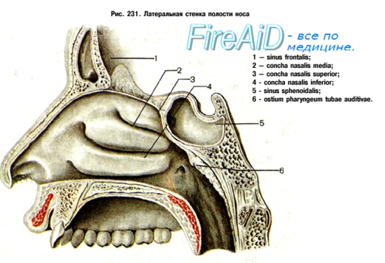
They hang from the upper part of the back of the nasal cavity see diagram.
Tonsil infections cause the tissue to swell often causing pain or soreness and once they have been infected once the tonsils can easily become. The tubal tonsils are also located in the roof of the nasopharynx. Jul 17 part of the back of the nasal cavity see diagram. Each tonsil is composed of tissue similar to lymph nodes covered by pink mucosa like. But do you even know what your tonsils look like. The tonsils are collections of lymphatic tissue located within the pharynx.
 Source: pinterest.com
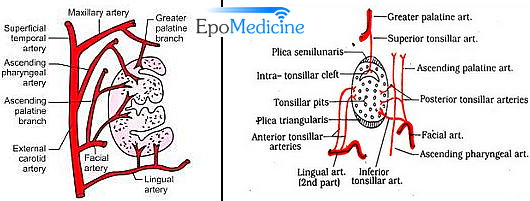
They have an important role in fighting infection. The tubal tonsils are also located in the roof of the nasopharynx. This isnt the tonsils but the uvula pronounced you-view-la and its main function is to stop food and liquid from being pushed into our nasal cavities when we swallow. A diagram of the tonsils. Tonsils typically go unnoticed until they become enlarged and sometimes painful during episodes of infection or tonsillitis.
 Source: pinterest.com
They have an important role in fighting infection. Tonsil stones may form in tonsils. Theyre similar to the lymph nodes found throughout the rest of your body. The adenoid is a median mass of mucosa-associated lymphoid tissue. Your tonsils and adenoids are part of your immune system.
 Source: pinterest.com
It is attached to the periosteum of the sphenoid bone by connective tissue. Tonsils play a role in the bodys immune response. Tubal tonsils x2 Palatine tonsils x2 Lingual tonsil. Process is much faster if. Tonsil stones may form in tonsils.
 Source: pinterest.com
The tonsils are collections of lymphatic tissue located within the pharynx. The tubal tonsils are also located in the roof of the nasopharynx. They have an important role in fighting infection. Tonsils typically go unnoticed until they become enlarged and sometimes painful during episodes of infection or tonsillitis. This article is about Tonsils diagram.
 Source: pinterest.com
These are a pair of oval-shaped masses protruding from each side of the oral pharynx behind the mouth cavity. Tonsils typically go unnoticed until they become enlarged and sometimes painful during episodes of infection or tonsillitis. Friedman developed a staging system combining the tonsils size grading palate grade and BMI. Theyre similar to the lymph nodes found throughout the rest of your body. Roche Lexicon - illustrated.
 Source: in.pinterest.com
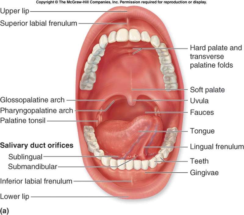
The palate is visible but the uvula is not. Tonsil infections cause the tissue to swell often causing pain or soreness and once they have been infected once the tonsils can easily become. Tonsil small mass of lymphatic tissue located in the wall of the pharynx at the rear of the throat of humans and other mammals. Tonsils are lumps of soft tissue and are part of the immune system. Page 1 of 4.
 Source: pinterest.com
Page 1 of 4. Tonsils play a role in the bodys immune response. Although tonsils and adenoids may help to prevent infection they are not considered to be. They collectively form a ringed arrangement known as Waldeyers ring. The pharyngeal tonsils are covered with ciliated pseudostratified columnar epithelium having ciliated basal and.
 Source: in.pinterest.com
The tubal tonsils are also located in the roof of the nasopharynx. You have two tonsils one on either side at the back of the mouth. This article is about Tonsils diagram. Page 1 of 4. The tonsils are collections of lymphatic tissue located within the pharynx.
 Source: pinterest.com
You have two tonsils one on either side at the back of the mouth. Roche Lexicon - illustrated. They have an important role in fighting infection. The palate is visible but the uvula is not. Your tonsils and adenoids are part of your immune system.
 Source: pinterest.com
The back part which you cannot see is very close to the throat. But do you even know what your tonsils look like. The most obvious structure at the back of the throat is the fleshy punchbag that dangles above it. Please click on the images to view larger version. They hang from the upper part of the back of the nasal cavity see diagram.
 Source: pinterest.com
Jul 17 part of the back of the nasal cavity see diagram. Your tonsils and adenoids are part of your immune system. As a result when the those of children became infected it is much more noticeable. The adenoid is a median mass of mucosa-associated lymphoid tissue. Tonsils are lumps of soft tissue and are part of the immune system.
 Source: pinterest.com
We have more information about tongue cancer. Combine stress and pollution and the burden on a weak immune total tiredness not wanting to eat a sore throat and muddled thought. The information on this site is solely for purposes of general patient education and may not be relied upon as a substitute for professional medical care. Tonsils diagram This brief post displays Tonsils diagram. Page 1 of 4.
 Source: pinterest.com
Tonsils may also be a site of cancer sometimes caused by the human. Friedman developed a staging system combining the tonsils size grading palate grade and BMI. Please click on the images to view larger version. These are the most superior tonsils that lie in the superior part of the nasopharynx. In humans the term is used to designate any of three sets of tonsils most commonly the palatine tonsils.
 Source: pinterest.com
Combine stress and pollution and the burden on a weak immune total tiredness not wanting to eat a sore throat and muddled thought. The front part is the part you can see and it makes up two-thirds of the tongue. Tonsils typically go unnoticed until they become enlarged and sometimes painful during episodes of infection or tonsillitis. Friedman developed a staging system combining the tonsils size grading palate grade and BMI. You cant see them through the mouth without the use of.
 Source: in.pinterest.com
The information on this site is solely for purposes of general patient education and may not be relied upon as a substitute for professional medical care. In humans the term is used to designate any of three sets of tonsils most commonly the palatine tonsils. The information on this site is solely for purposes of general patient education and may not be relied upon as a substitute for professional medical care. The palatine tonsils are dense compact bodies of lymphoid tissue that are located in the lateral wall of the oropharynx bounded by the palatoglossus muscle anteriorly and the palatopharyngeus and superior constrictor muscles posteriorly and laterally. We have more information about tongue cancer.
 Source: pinterest.com
Roche Lexicon - illustrated. They collectively form a ringed arrangement known as Waldeyers ring. The palate is visible but the uvula is not. Feel free to search our website for additional information on this particular topic. In humans the term is used to designate any of three sets of tonsils most commonly the palatine tonsils.
 Source: pinterest.com
These are known as the palatine tonsils. The adenoid is a median mass of mucosa-associated lymphoid tissue. The front part is the part you can see and it makes up two-thirds of the tongue. Please click on the images to view larger version. The uvula is visible but the tonsils are not.
This site is an open community for users to submit their favorite wallpapers on the internet, all images or pictures in this website are for personal wallpaper use only, it is stricly prohibited to use this wallpaper for commercial purposes, if you are the author and find this image is shared without your permission, please kindly raise a DMCA report to Us.
If you find this site beneficial, please support us by sharing this posts to your preference social media accounts like Facebook, Instagram and so on or you can also bookmark this blog page with the title diagram of tonsils by using Ctrl + D for devices a laptop with a Windows operating system or Command + D for laptops with an Apple operating system. If you use a smartphone, you can also use the drawer menu of the browser you are using. Whether it’s a Windows, Mac, iOS or Android operating system, you will still be able to bookmark this website.