Ecg diagram explanation
Home » Wallpapers » Ecg diagram explanationYour Ecg diagram explanation images are available. Ecg diagram explanation are a topic that is being searched for and liked by netizens now. You can Find and Download the Ecg diagram explanation files here. Download all free photos and vectors.
If you’re looking for ecg diagram explanation pictures information related to the ecg diagram explanation topic, you have visit the ideal site. Our site always gives you suggestions for seeing the highest quality video and picture content, please kindly search and locate more informative video articles and graphics that match your interests.
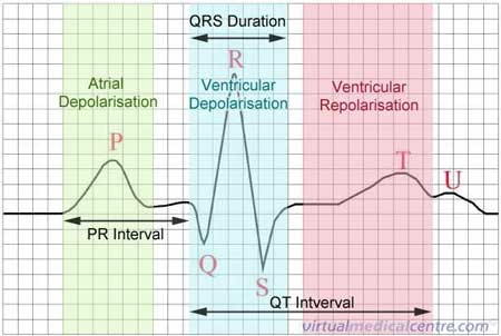
Ecg Diagram Explanation. The electrocardiograph presents these electrical events in a diagram referred to as electrocardiogram ECG. The main blocks of an ECG machine and the function of each block is explained below. An ECG machine records this activity via electrodes on the skin and displays it graphically. T wave usually same directionT wave usually same direction as QRS - ventricular repolarisation.
 Ecg Electrocardiogram Heartbeat Monitoring Information Myvmc From myvmc.com
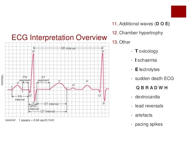
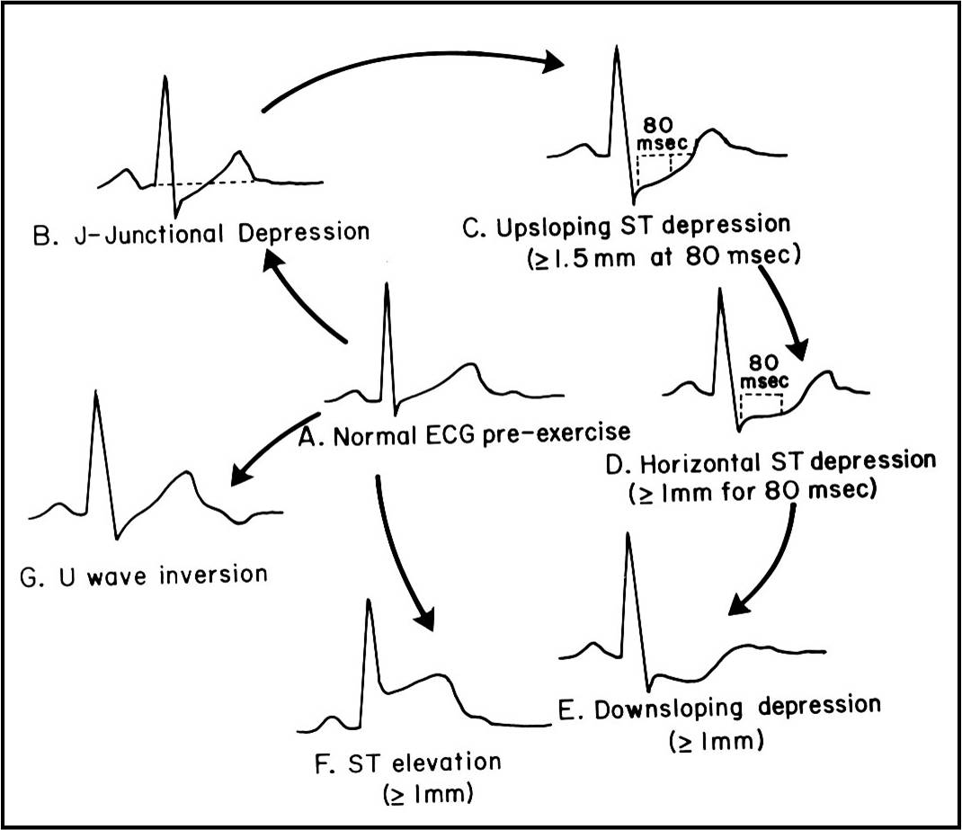
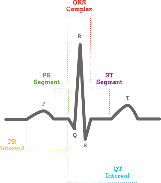
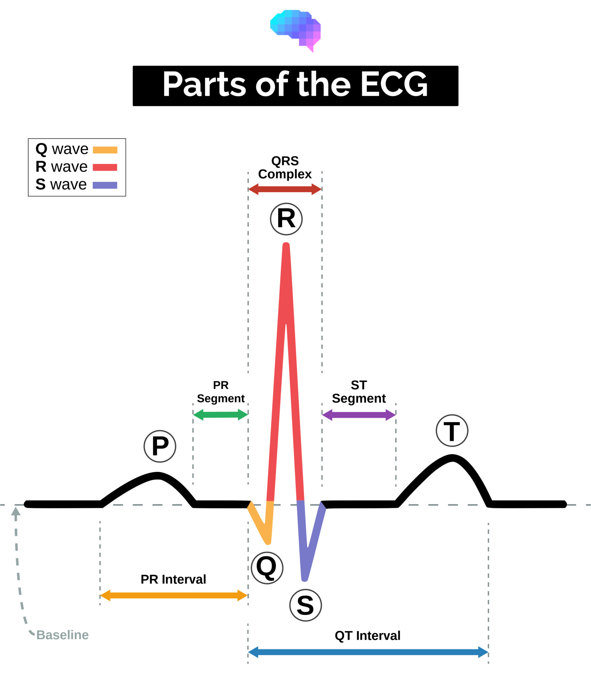
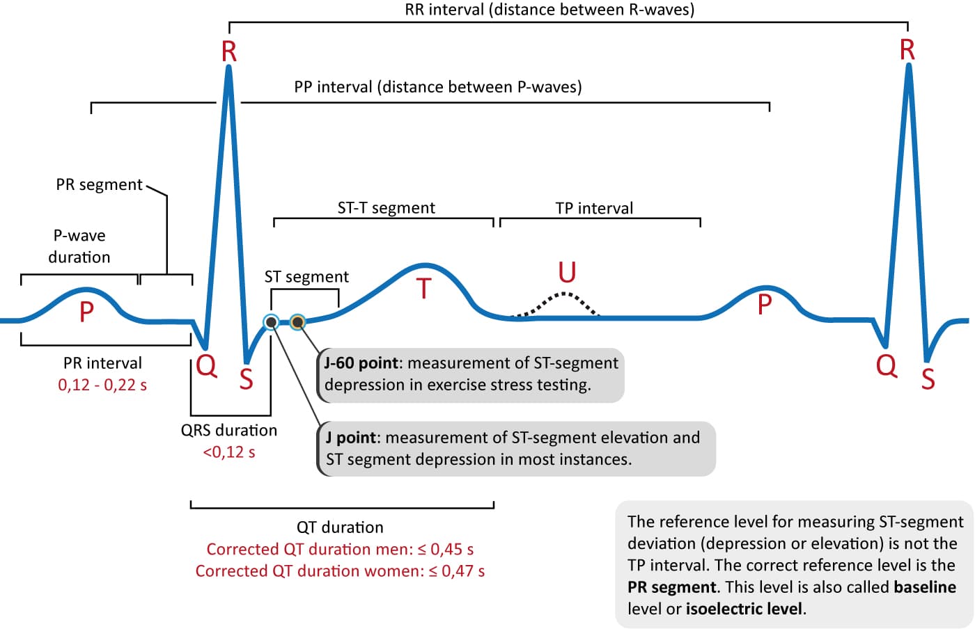
PR Interval impulse from atria to ventriclesto ventricles. A ladder diagram is a diagram to explain arrhythmias. QRS complex ventricular depolarisation. I IlI. ST segment isoelectric - part of repolarisation. One to each limb and six across the chest.
-30 to 90.
Middle of T wave. One to each limb and six across the chest. Provides information on atrial depolarization and the P wave ventricular depolarization a. An ECG machine records this activity via electrodes on the skin and displays it graphically. ST segment isoelectric - part of repolarisation. Before discussing each component in detail a brief overview of the waves and intervals is given.
 Source: researchgate.net
EKG or ECG waveform parts are explained clearly to make EKG interpretation easy. The electrocardiogram ECG or EKG is a noninvasive test that is used to reflect underlying heart conditions by measuring the electrical activity of the heart. Conversely a wave traveling away from the positive lead will inscribe a downward deflection. With vector manipulation ECG machine creates aVR aVL aVF. The main blocks of an ECG machine and the function of each block is explained below.
 Source: commons.wikimedia.org
Draw the Electrical Cardiac Cycle Events ECG. QRS complex ventricular depolarisation. Peak of R wave. ECG interpretation includes an assessment of the morphology appearance of the waves and intervals on the ECG curve. Conversely a wave traveling away from the positive lead will inscribe a downward deflection.
 Source: slideshare.net
P wave atrial depolarisation. PR Interval impulse from atria to ventriclesto ventricles. Lead I is calculated using data from the electrodes on both the right and left arm. -30 to 90. Augmented Limb Leads Unipolar.
 Source: pinterest.com
The EKG waveform comes from a measurement of surface voltages between 2 leads. The main blocks of an ECG machine and the function of each block is explained below. Lead I is calculated using data from the electrodes on both the right and left arm. The EKG waveform comes from a measurement of surface voltages between 2 leads. By positioning leads electrical sensing devices on the body in standardized locations information about many heart conditions can be learned by looking for characteristic patterns on the ECG.
 Source: youtube.com
Many medical students and even some more experienced medical professionals struggle with ECG interpretation. The electrocardiogram ECG or EKG is a noninvasive test that is used to reflect underlying heart conditions by measuring the electrical activity of the heart. Right Axis Deviation -RVH -Left posterior hemiblock -Dextrocardia -Ectopic ventricular beats and. T wave usually same directionT wave usually same direction as QRS - ventricular repolarisation. The electrocardiograph presents these electrical events in a diagram referred to as electrocardiogram ECG.
 Source: researchgate.net
The origin of impulse formation sinus node for the first two beats and AV junction for the third beat and the conduction in the heart are shown. Middle of T wave. QT Interval This intervalQT Interval This interval spans. The figure shows a simple ladder diagram for normal sinus rhythm followed by av-nodal extrasystole. Learn the seven steps to interpret an ECG with help of an analysis algorithm and test your knowledge by taking the ECG quiz.
 Source: ecg.utah.edu
Peak of R wave. An ECG machine records this activity via electrodes on the skin and displays it graphically. Hexaxial System Used to determine electrical axis What is the normal axis for the heart. Corresponds to beginning of ventricular diastole. Peak of R wave.
 Source: researchgate.net
An ECG electrode is a conductive pad that is attached to the skin to record electrical activity. T wave usually same directionT wave usually same direction as QRS - ventricular repolarisation. Working principle normal ECG wave application of ECG August 15 2018 Gaurab Karki Anatomy and Physiology Zoology 0 Electrocardiogram refers to the recording of electrical changes that occurs in heart during a cardiac cycle. Therefore ECG interpretation requires a structured assessment of the waves and intervals. Many medical students and even some more experienced medical professionals struggle with ECG interpretation.
 Source: youtube.com
QT Interval This intervalQT Interval This interval spans. Corresponds to beginning of ventricular systole. Many medical students and even some more experienced medical professionals struggle with ECG interpretation. ECG Machine Block diagram and working. Conversely a wave traveling away from the positive lead will inscribe a downward deflection.
 Source: alivecor.com
ST segment isoelectric - part of repolarisation. Therefore ECG interpretation requires a structured assessment of the waves and intervals. -30 to 90. Many medical students and even some more experienced medical professionals struggle with ECG interpretation. Heart rate heart rhythm electrical heart axis the pr interval repolarization.
 Source: brainkart.com
An ECG electrode is a conductive pad that is attached to the skin to record electrical activity. Hexaxial System Used to determine electrical axis What is the normal axis for the heart. Corresponds to beginning of ventricular systole. The data gathered from these electrodes allows the 12 leads of the ECG to be calculated eg. With vector manipulation ECG machine creates aVR aVL aVF.
 Source: examfear.com
An ECG involves attaching 10 electrical cables to the body. T wave usually same directionT wave usually same direction as QRS - ventricular repolarisation. Hexaxial System Used to determine electrical axis What is the normal axis for the heart. Augmented Limb Leads Unipolar. -30 to 90.
 Source: myvmc.com
Always start with the ECG. Corresponds to beginning of ventricular systole. Hexaxial System Used to determine electrical axis What is the normal axis for the heart. Learn the seven steps to interpret an ECG with help of an analysis algorithm and test your knowledge by taking the ECG quiz. An ECG involves attaching 10 electrical cables to the body.
 Source: geekymedics.com
We know the ECG electrodes mainly used for the pickup of ECG are five in number. Conversely a wave traveling away from the positive lead will inscribe a downward deflection. EKG or ECG waveform parts are explained clearly to make EKG interpretation easy. Therefore ECG interpretation requires a structured assessment of the waves and intervals. Draw the Electrical Cardiac Cycle Events ECG.
 Source: ecgwaves.com
Learn the seven steps to interpret an ECG with help of an analysis algorithm and test your knowledge by taking the ECG quiz. The other end of electrode passes through defibrillator protection circuit. The Standard Limb Leads are used to display a graph of the potential difference recorded between two limbs at a time ergo they are bipolar. The one end of the electrode leads are connected along RA LA chest and LL of the patient. Learn the seven steps to interpret an ECG with help of an analysis algorithm and test your knowledge by taking the ECG quiz.
 Source: researchgate.net
Therefore ECG interpretation requires a structured assessment of the waves and intervals. Standard Limb Leads Bipolar. This is the principle behind the ECG. In these leads one limb carries a positive electrode and the other. Heart rate heart rhythm electrical heart axis the pr interval repolarization.
 Source: ib.bioninja.com.au
Standard Limb Leads Bipolar. Corresponds to beginning of ventricular systole. Learn the seven steps to interpret an ECG with help of an analysis algorithm and test your knowledge by taking the ECG quiz. An ECG electrode is a conductive pad that is attached to the skin to record electrical activity. P wave atrial depolarisation.
 Source: nursingofficer.net
Middle of T wave. T wave usually same directionT wave usually same direction as QRS - ventricular repolarisation. Peak of R wave. Middle of T wave. The electrocardiograph presents these electrical events in a diagram referred to as electrocardiogram ECG.
This site is an open community for users to do submittion their favorite wallpapers on the internet, all images or pictures in this website are for personal wallpaper use only, it is stricly prohibited to use this wallpaper for commercial purposes, if you are the author and find this image is shared without your permission, please kindly raise a DMCA report to Us.
If you find this site value, please support us by sharing this posts to your preference social media accounts like Facebook, Instagram and so on or you can also save this blog page with the title ecg diagram explanation by using Ctrl + D for devices a laptop with a Windows operating system or Command + D for laptops with an Apple operating system. If you use a smartphone, you can also use the drawer menu of the browser you are using. Whether it’s a Windows, Mac, iOS or Android operating system, you will still be able to bookmark this website.