Ecg lead diagram
Home » Background » Ecg lead diagramYour Ecg lead diagram images are ready in this website. Ecg lead diagram are a topic that is being searched for and liked by netizens now. You can Get the Ecg lead diagram files here. Find and Download all royalty-free images.
If you’re searching for ecg lead diagram pictures information related to the ecg lead diagram interest, you have visit the ideal site. Our site frequently gives you hints for seeing the maximum quality video and picture content, please kindly surf and find more informative video articles and graphics that match your interests.
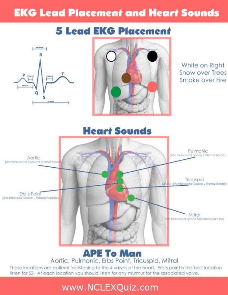
Ecg Lead Diagram. The main blocks of an ECG machine and the function of each block is explained below. You can view these and other helpful diagrams1. A 5-Lead ECG uses 4 limb leads and 1 chest lead. When viewing the EKG strip V4-V6 on the strip will be referred to as V-13-15.
 The Ecg Leads Electrodes Limb Leads Chest Precordial Leads 12 Lead Ecg Ekg Ecg Learning Electrodes Ekg Ecg Interpretation From pinterest.com
The electrical signals from the heart are measured with surface electrodes. This application note will attempt to give the reader a. 11102010 111933 AM. Three 3 lead ECG ElectrodeCable Placement. The Standard 12 Lead ECG. The electrical activity of this lead is measured and recorded as part of the ECG.
3 lead ECG cable Placement there are two ways Way 1.
When viewing the EKG strip V4-V6 on the strip will be referred to as V-13-15. The electrical activity of this lead is measured and recorded as part of the ECG. 3-Lead ECG A 3-Lead ECG uses 3 electrodes that are labeled white black and red. The electrical signals from the heart are measured with surface electrodes. Additional Lead placements Right sided ECG electrode placement. There are several approaches to recording a right-sided ECG.
 Source: id.pinterest.com
One electrode adjacent each clavicle bone on the upper chest and a third electrode adjacent the patients lower left abdomen. The three types of electrode systems are. 4th intercostal space right sternal border. A complete set of right-sided leads is obtained by placing leads V1-6 in a mirror-image position on the right side of the chest see diagram below. On a standard 12-lead EKG there are only 10 electrodes which are listed in the table below.
 Source: pt.pinterest.com
Monitors one of the three leads. ECG Machine Block diagram and working. These are present on the basic 3-lead monitors and also on the 12-lead EKG machines. Standard Limb Leads Bipolar. Three Lead analysis Lead I Lead II and aVF.
 Source: pinterest.com
The protection circuit has buffer amplifier and over-load voltage protection circuit. The other end of electrode passes through defibrillator protection circuit. Breast tissue can impact on the ECG amplitude due to the increased distance between the electrode and the heart when ECG electrodes are placed over the chest Rautaharuju et. 4th intercostal space left sternal border. The one end of the electrode leads are connected along RA LA chest and LL of the patient.
 Source: pinterest.com
4th intercostal space right sternal border. Additional Lead placements Right sided ECG electrode placement. ECG limb lead placement diagram. An ECG lead is a graphical representation of the hearts electrical activity which is calculated by analysing data from several ECG electrodes. By placing these electrodes at appropriate parts of.
 Source: pinterest.com
This application note will attempt to give the reader a. This placement takes place on both the right. A 5-Lead ECG uses 4 limb leads and 1 chest lead. A 3-lead configuration requires the placement of three electrodes. Limb Lead Placement Diagram The limb electrodes can be far down on the limbs or close to the hipsshoulders as long as they are placed symmetrically.
 Source: pinterest.com
The placement procedure is pretty standard with the Leads being placed on both the left and right arms and the legs as well. It is Typical Bipolar Lead form and Monitor reads as Lead III III. ECG Lead System Configuration. The one end of the electrode leads are connected along RA LA chest and LL of the patient. The electrical activity of this lead is measured and recorded as part of the ECG.
 Source: pinterest.com
ECG Lead System Configuration. You can view these and other helpful diagrams1. The one end of the electrode leads are connected along RA LA chest and LL of the patient. AVR aVL aVF. Normally we use the above set up for the measurement and plotting of ECG.
 Source: pinterest.com
How the 12 lead ECG works. The electrical signals from the heart are measured with surface electrodes. Monitors one of the three leads. Midway between leads V2 and V4. AVR aVL aVF.
 Source: pinterest.com
The Standard 12 Lead ECG. One electrode adjacent each clavicle bone on the upper chest and a third electrode adjacent the patients lower left abdomen. 5th intercostal space anterior axillary line. When interpreted accurately an ECG can detect and monitor a host of heart conditions from arrhythmias to coronary heart disease to electrolyte imbalance. We know the ECG electrodes mainly used for the pickup of ECG are five in number.
 Source: pinterest.com
5th intercostal space anterior axillary line. Positive and negative poles for leads I II III In physics two vectors leads are equal as long as they are parallel and same polarity Move the leads to pass through the center of the heart With vector manipulation ECG machine creates aVR aVL aVF. You can view these and other helpful diagrams1. The basic leads consist of leads I II and III and the augmented leads AVR AVL and AVF. On a standard 12-lead EKG there are only 10 electrodes which are listed in the table below.
 Source: pinterest.com
3-Lead ECG A 3-Lead ECG uses 3 electrodes that are labeled white black and red. There are several complementary approaches to estimating QRS axis which are summarized below. 12-Lead ECG Placement Guide with Illustrations. By placing these electrodes at appropriate parts of. The basic leads consist of leads I II and III and the augmented leads AVR AVL and AVF.
 Source: pinterest.com
Monitors one of the three leads. Electrocardiography ECG is the interpretation of the electrical activity of ones heart over a period of time. Three 3 lead ECG ElectrodeCable Placement. The basic leads consist of leads I II and III and the augmented leads AVR AVL and AVF. This lead position should be avoided as it may result in distorted ECG morphology it may alter ST.
 Source: ar.pinterest.com
There are several approaches to recording a right-sided ECG. To clarify leads will equal. This application note will attempt to give the reader a. A 12-lead ECG records 12 of these leads producing 12 separate graphs. ECG Lead System Configuration.
 Source: br.pinterest.com
Microsoft PowerPoint - CAR-205 Basic 12 lead EKG v4ppt Author. I IlI III. A 12-lead ECG records 12 of these leads producing 12 separate graphs. There are several approaches to recording a right-sided ECG. The three types of electrode systems are.
 Source: pinterest.com
Lead refers to an imaginary line between two ECG electrodes. There are several complementary approaches to estimating QRS axis which are summarized below. A 12-lead ECG records 12 leads. This section describes the basic components of the ECG and the lead system used to record the ECG tracings. To clarify leads will equal.
 Source: pinterest.com
Understanding the 12-lead ECG. The ECG Leads Polarity and Einthovens Triangle. Three Lead analysis Lead I Lead II and aVF. 4th intercostal space left sternal border. Midway between leads V2 and V4.
 Source: pinterest.com
How the 12 lead ECG works. ECG Recording Setup Block Diagram. The placement procedure is pretty standard with the Leads being placed on both the left and right arms and the legs as well. There are several complementary approaches to estimating QRS axis which are summarized below. Placed the red electrode within the frame of rib cageright under the clavicle near shoulder see chart in follow picture LA.
 Source: pinterest.com
The Quadrant Method Lead I and aVF. Another pair of the ECGs electrodes are then set between the fourth and fifth ribs respectively. Electrocardiography ECG is the interpretation of the electrical activity of ones heart over a period of time. 4th intercostal space left sternal border. On a standard 12-lead EKG there are only 10 electrodes which are listed in the table below.
This site is an open community for users to do sharing their favorite wallpapers on the internet, all images or pictures in this website are for personal wallpaper use only, it is stricly prohibited to use this wallpaper for commercial purposes, if you are the author and find this image is shared without your permission, please kindly raise a DMCA report to Us.
If you find this site good, please support us by sharing this posts to your own social media accounts like Facebook, Instagram and so on or you can also save this blog page with the title ecg lead diagram by using Ctrl + D for devices a laptop with a Windows operating system or Command + D for laptops with an Apple operating system. If you use a smartphone, you can also use the drawer menu of the browser you are using. Whether it’s a Windows, Mac, iOS or Android operating system, you will still be able to bookmark this website.