Epidermis diagram
Home » Background » Epidermis diagramYour Epidermis diagram images are ready. Epidermis diagram are a topic that is being searched for and liked by netizens today. You can Find and Download the Epidermis diagram files here. Download all royalty-free vectors.
If you’re looking for epidermis diagram images information related to the epidermis diagram interest, you have visit the ideal site. Our site always provides you with hints for seeing the maximum quality video and picture content, please kindly search and find more informative video content and graphics that fit your interests.
Epidermis Diagram. Learn vocabulary terms and more with flashcards games and other study tools. INTEGUMENTARY SYSTEM PART III. Epidermis Cell Structure Aesthetic Dermatology Skin Cells Skin Skin Structure Diagram Human Anatomy And Physiology Physiology More information. The skins structure is made up of an intricate network which serves as the bodys initial barrier against pathogens UV light and chemicals and mechanical injury.
 Anatomi Dan Fisiologi Kulit Skin Anatomy Integumentary System Anatomy From id.pinterest.com
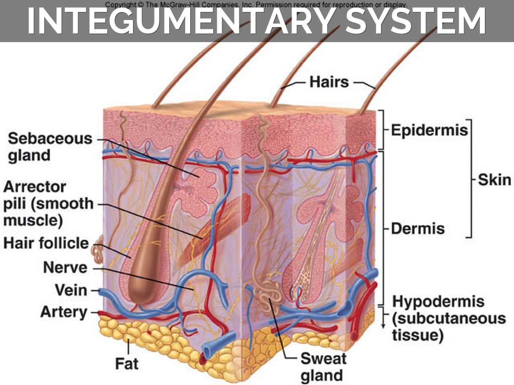
How many rows of cells are in epidermis. Skin is the largest organ in the body and covers the bodys entire external surface. The upper layer of skin composed of the Stratum Corneum stratum Lucidum Stratum Granulosum Stratum Spinosum and Stratum Germinativum. This skin diagram clearly shows all the layers of skin. Three Basic Cell Types in the Epidermis The three basic cell types in the epidermis include keratinocytes some labeled K and Langerhans cells L in the Malpighian layer and melanocytes M in the basal layer. Learn vocabulary terms and more with flashcards games and other study tools.
The epidermis usually consists of a single layer of cells which cover the whole outer surface of the plant body.
It is a continuous layer except for certain small pores called stomata and lenticels. We will now go over the skins layers in more detail. There are no blood vessels in the epidermis but. Are made of epithelial tissue part of epidermis are located in dermis project through the skin surface The Hair Follicle Is located deep in dermis made of epithleial tissue. How many rows of cells are in epidermis. Epidermis dermis fat cells hair shaft hair follicle hair erector muscle sweat gland pore of sweat gland sebaceous gland blood.
 Source: pinterest.com
The dermis the epidermis. The epidermis layer provides a barrier to infection from environmental pathogens and regulates the amount of water released from the body into the atmosphere through transepidermal water loss. The dermis is the layer of the skin present beneath the epidermis of the skin. Psoriasis in section skin with detailed epidermis in stratum corneum granular and germinative dermis and hypodermis. This diagram shows schematically the four different layers found in the epidermis of most skin thin skin.
 Source: pinterest.com
The outermost layer of the skin is. The stratum basale also called the stratum germinativum is the deepest epidermal layer and attaches the epidermis to the basal lamina below which lie the layers of the dermis. This skin diagram clearly shows all the layers of skin. The epidermis is a dynamic structure acting as a semi-permeable barrier with a layer of flat anuclear cells at the surface stratum corneum. Layers in the Epidermis.
 Source: pinterest.com
The word is derived from two words of Greek origin epi upon and derma skin. There are no blood vessels in the epidermis but. INTEGUMENTARY SYSTEM PART III. The epidermis usually consists of a single layer of cells which cover the whole outer surface of the plant body. The dermis the epidermis fat layer 2.
 Source: pinterest.com
Table of contentswhat is a fishbone diagramhowread more free fishbone diagram templates word excel pdf This ielts diagram task 1guide will get you to learn the following aspects of academic writing task 1. The epidermis layer provides a barrier to infection from environmental pathogens and regulates the amount of water released from the body into the atmosphere through transepidermal water loss. Three Basic Cell Types in the Epidermis The three basic cell types in the epidermis include keratinocytes some labeled K and Langerhans cells L in the Malpighian layer and melanocytes M in the basal layer. Cells divide in the basal layer and move up through the layers above changing their appearance as they move from one layer to the next. Epidermis Cell Structure Aesthetic Dermatology Skin Cells Skin Skin Structure Diagram Human Anatomy And Physiology Physiology More information.
 Source: id.pinterest.com
The epidermis is a dynamic structure acting as a semi-permeable barrier with a layer of flat anuclear cells at the surface stratum corneum. The epidermis is a dynamic structure acting as a semi-permeable barrier with a layer of flat anuclear cells at the surface stratum corneum. It is in direct contact with. In this article we will discuss about the structure of epidermis in plants. The stratum basale also called the stratum germinativum is the deepest epidermal layer and attaches the epidermis to the basal lamina below which lie the layers of the dermis.
 Source: pinterest.com
The epidermis is the outermost layer of the skin which is composed of cells called keratinocytes made of a protein called keratin. In this article we will discuss about the structure of epidermis in plants. The epidermis is composed of multiple layers of flattened cells that overlie. It is made up of three layers the epidermis dermis and the hypodermis all three of which vary significantly in their anatomy and function. As can be seen in the skin diagram the outermost layer of the skin is called the epidermis layer.
 Source: pinterest.com
The skins structure is made up of an intricate network which serves as the bodys initial barrier against pathogens UV light and chemicals and mechanical injury. The epidermis is the outermost of the three layers that make up the skin the inner layers being the dermis and hypodermis. Diagram of the skin and hair includes sweat gland Skin. The epidermis is composed of multiple layers of flattened cells that overlie. The word is derived from two words of Greek origin epi upon and derma skin.
 Source: pinterest.com
Table of contentswhat is a fishbone diagramhowread more free fishbone diagram templates word excel pdf This ielts diagram task 1guide will get you to learn the following aspects of academic writing task 1. The epidermis usually consists of a single layer of cells which cover the whole outer surface of the plant body. Are made of epithelial tissue part of epidermis are located in dermis project through the skin surface The Hair Follicle Is located deep in dermis made of epithleial tissue. The dermis the epidermis fat layer 2. We will now go over the skins layers in more detail.
 Source: pinterest.com
Tissue creating an external covering of the body. How many rows of cells are in epidermis. The epidermis is the outermost of the three layers that make up the skin the inner layers being the dermis and hypodermis. ACCESSORY STRUCTURES Integumentary Accessory Structures Hair hair follicles sebaceous glands sweat glands and nails. Cells divide in the basal layer and move up through the layers above changing their appearance as they move from one layer to the next.
 Source: pinterest.com
Integumentary system quiz and answers. This skin diagram clearly shows all the layers of skin. Start studying 32 Epidermis. Which is the thickest layer. One of the best ways to start learning about a new system organ or region is with a labeled diagram showing you all of the main structures found within it.
 Source: pinterest.com
The upper layer of skin composed of the Stratum Corneum stratum Lucidum Stratum Granulosum Stratum Spinosum and Stratum Germinativum. Start studying 32 Epidermis. The dermis the epidermis. Diagram of the skin and hair includes sweat gland Skin. The skins structure is made up of an intricate network which serves as the bodys initial barrier against pathogens UV light and chemicals and mechanical injury.
 Source: pinterest.com
The dermis is the layer of the skin present beneath the epidermis of the skin. The word is derived from two words of Greek origin epi upon and derma skin. The epidermis layer provides a barrier to infection from environmental pathogens and regulates the amount of water released from the body into the atmosphere through transepidermal water loss. The epidermis usually consists of a single layer of cells which cover the whole outer surface of the plant body. Three Basic Cell Types in the Epidermis The three basic cell types in the epidermis include keratinocytes some labeled K and Langerhans cells L in the Malpighian layer and melanocytes M in the basal layer.
 Source: pinterest.com
The word is derived from two words of Greek origin epi upon and derma skin. Diagram of the skin and hair includes sweat gland Skin. Epidermis Cell Structure Aesthetic Dermatology Skin Cells Skin Skin Structure Diagram Human Anatomy And Physiology Physiology More information. The epidermis layer provides a barrier to infection from environmental pathogens and regulates the amount of water released from the body into the atmosphere through transepidermal water loss. We will now go over the skins layers in more detail.
 Source: pinterest.com
The word is derived from two words of Greek origin epi upon and derma skin. How many rows of cells are in epidermis. Skin is the largest organ in the body and covers the bodys entire external surface. Three Basic Cell Types in the Epidermis The three basic cell types in the epidermis include keratinocytes some labeled K and Langerhans cells L in the Malpighian layer and melanocytes M in the basal layer. The epidermis is made of four main layers and functions by protecting and safeguarding the internal cells and tissues.
 Source: pinterest.com
The epidermis regenerates in orderly fashion by cell division of keratinocytes in the basal layer with maturing daughter cells becoming increasingly keratinised as they move to the skin surface. The epidermis regenerates in orderly fashion by cell division of keratinocytes in the basal layer with maturing daughter cells becoming increasingly keratinised as they move to the skin surface. As can be seen in the skin diagram the outermost layer of the skin is called the epidermis layer. The stratum basale also called the stratum germinativum is the deepest epidermal layer and attaches the epidermis to the basal lamina below which lie the layers of the dermis. Learn vocabulary terms and more with flashcards games and other study tools.
 Source: pinterest.com
This skin diagram clearly shows all the layers of skin. Start studying 32 Epidermis. Add the following labels to the diagram of the skin shown below. This will also help you to draw the structure and diagram of epidermis in plants. Are made of epithelial tissue part of epidermis are located in dermis project through the skin surface The Hair Follicle Is located deep in dermis made of epithleial tissue.
 Source: pinterest.com
This epidermis of skin is a keratinized stratified squamous epithelium. The epidermis layer provides a barrier to infection from environmental pathogens and regulates the amount of water released from the body into the atmosphere through transepidermal water loss. One of the best ways to start learning about a new system organ or region is with a labeled diagram showing you all of the main structures found within it. This epidermis of skin is a keratinized stratified squamous epithelium. It is made up of three layers the epidermis dermis and the hypodermis all three of which vary significantly in their anatomy and function.
 Source: pinterest.com
The epidermis is composed of multiple layers of flattened cells that overlie. The skins structure is made up of an intricate network which serves as the bodys initial barrier against pathogens UV light and chemicals and mechanical injury. The epidermis layer provides a barrier to infection from environmental pathogens and regulates the amount of water released from the body into the atmosphere through transepidermal water loss. Table of contentswhat is a fishbone diagramhowread more free fishbone diagram templates word excel pdf This ielts diagram task 1guide will get you to learn the following aspects of academic writing task 1. The outermost layer of skin consisting of dead and Keratinization cells.
This site is an open community for users to submit their favorite wallpapers on the internet, all images or pictures in this website are for personal wallpaper use only, it is stricly prohibited to use this wallpaper for commercial purposes, if you are the author and find this image is shared without your permission, please kindly raise a DMCA report to Us.
If you find this site value, please support us by sharing this posts to your preference social media accounts like Facebook, Instagram and so on or you can also save this blog page with the title epidermis diagram by using Ctrl + D for devices a laptop with a Windows operating system or Command + D for laptops with an Apple operating system. If you use a smartphone, you can also use the drawer menu of the browser you are using. Whether it’s a Windows, Mac, iOS or Android operating system, you will still be able to bookmark this website.